Overview of Dentitions (Part A)
Occlusion and Malocclusion
Occlusion is the relationship between the maxillary and mandibular teeth when the upper and lower jaws are fully closed. Occlusion-related problems could affect the teeth, joints, and muscles of the head and neck.
Malocclusion refers to abnormal or malpositioned relationships of maxillary teeth to mandibular teeth when they are in centric occlusion. Malocclusion can also cause periodontal disease.
Centric and Functional Occlusion
Centric occlusion occurs when the jaws are in a position that produces maximal stable contact between the occluding surfaces of the maxillary and mandibular teeth. Centric occlusion serves as the standard for normal occlusion. It occurs when jaws are closed in a position that produces maximal stable contact between occluding surfaces of maxillary and mandibular teeth.
Functional occlusion is the term used to describe the teeth during biting and chewing movements.
Factors Affecting Occlusion
Altering the relationships between the teeth in the same or opposite arches can lead to malocclusion or abnormal occlusion. Some of the factors that can affect occlusion may include:
- Skeletofacial growth and development patterns in babies, children, and adolescents
- Abnormally large or small teeth
- Thumb-sucking habits
- Improper swallowing habits
- Premature loss of primary or permanent dentition
Angle’s Classification
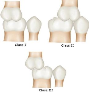
Dr. Edward H. Angle created Angle’s Classification system to describe and classify occlusion and malocclusion in adult dentition. The basis of this system is the relationship between the maxillary first molar and the mandibular first molar as viewed from the patient’s profile. Angle’s system assumes that the patient is occluding in the centric position.
To determine the occlusion classification, the oral health professional will often ask the patient to “bite together” while retracting the cheek to view the facial aspects of these two teeth.
 |
 |
| Close-up image of a human mouth showing teeth with dental malocclusion, specifically Angle’s Class 1, where the teeth are misaligned but the dental arches correctly meet. | Illustration depicting three different classes of teeth alignment according to Angle’s classification: Class I with normal occlusion, Class II with overbite, and Class III with underbite. |
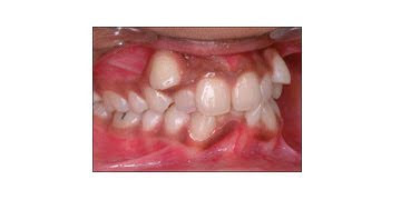
Class I
In Class I or neutroclusion, an ideal relationship exists between the jaws and the dental arches. The mesiobuccal cusp of the permanent maxillary first molar occludes (or aligns) with the mesiobuccal groove of the mandibular first molar.
Class II
In Class II or distoclusion, the mandibular arch is in a distal relationship to the maxillary arch. The mesiobuccal cusp of the permanent maxillary first molar occludes with the mesiobuccal groove of the permanent mandibular first molar. This often gives the appearance of protrusion of the maxillary anterior teeth over the mandibular anterior teeth.
The major group of Class II malocclusion has two subgroups – division 1 and division 2. These subgroups are based on the anterior teeth’ position, the palate’s shape, and the facial profile.
Class II, Division 1
Class II, Division 1: Lips are usually flat and parted, with the lower lip tucked behind the upper incisors. Maxillary anterior protrudes facially from the mandibular anterior, with a deep overbite (lab version).
 |
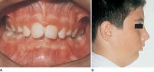 |
| Illustration of a human dental arch with a Class II Division 1 malocclusion, characterized by an overbite with upper teeth protruding significantly over the lower teeth. | Two images showing Class II Division 1 dental malocclusion: A) A frontal view of an open mouth displaying misaligned teeth, and B) A side profile of a young male with his eye area blacked out for privacy. |
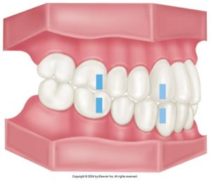
Class II, Division 2
Class II, Division 2: Maxillary incisors are not in labioversion. Maxillary central incisors are nearly normal anteroposteriorly, and they may be slightly in linguoversion. Maxillary central incisors are either upright or retruded, and lateral incisors are either tipped labially or overlap the central incisors with deep overbite.
 |
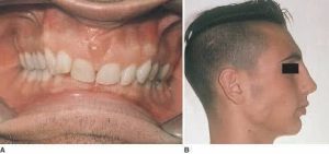 |
| Illustration of a human dental arch with a Class II Division 2 malocclusion, characterized by upper front teeth tilted inward while the back teeth maintain normal alignment. | Two images showing Class II Division 2 dental malocclusion: A) A frontal view of an open mouth displaying upper front teeth tilted inward, and B) A side profile of a young male with his eye area blacked out for privacy. |
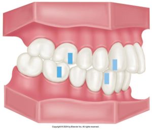
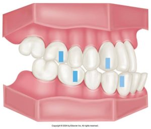
Class III
In Class III or mesioclusion, the mandibular arch is in a mesial relationship with the maxillary arch. The mesiobuccal cusp of the permanent maxillary first molar occludes distal to the mesiobuccal groove of the permanent mandibular first molar. This often gives the appearance of protrusion of the mandible.
 |
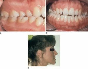 |
| Illustration of a human dental arch with Class III malocclusion, showing underbite where the lower teeth protrude past the upper teeth, with blue markers on specific teeth indicating areas of focus. | Three images illustrating Class III malocclusion: A) a Close-up of the lower jaw showing underbite, B) a Front view of an open mouth with lower teeth protruding beyond upper teeth, and C) a Side profile of a woman with her eye area blacked out to show the facial impact of the underbite. |
Activity 1: Occlusion
Stabilization of the Arches
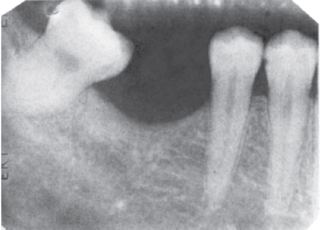
The image below shows a radiograph showing the mesial drift of the mandibular second molar after the first molar has been lost.
Closure
The dental arches remain stable and efficient in a healthy mouth with properly maintained dentition. Malocclusion or the loss of one or more teeth may greatly reduce the functioning and stability of the dentition.
Closure: Anterior teeth are not designed to support the occlusal forces on the entire dental arch fully; therefore, as the jaws close, the stronger posterior teeth come together first. The more delicate anterior teeth come together after the posterior teeth have assumed most of the load.
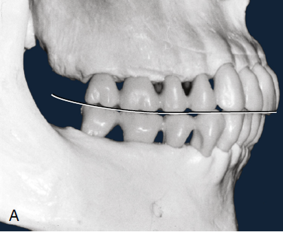
Curve of Spee
The occlusal surfaces of the posterior teeth do not form a flat plane. The teeth of the mandibular arch form a slightly curved plane, which appears concave (curved inward, like the inside of a bowl). The maxillary arch forms a curved plane that appears convex (curved outward, like the outside of a bowl). The curvature formed by the maxillary and mandibular arches in occlusion is the Spee curve. On the image of the radiograph below, the occlusal line of the teeth appears to be smiling.
 |
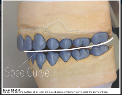 |
| 3D scan of a human skull showing the dental arch with teeth, featuring a line indicating the Curve of Spee, which highlights the curvature of the occlusal surfaces of the molars and premolars. | Model of a human dental arch displaying the Curve of Spee, with a line running across the occlusal surfaces of the teeth to illustrate the natural curvature in a side view of the dental cast. |
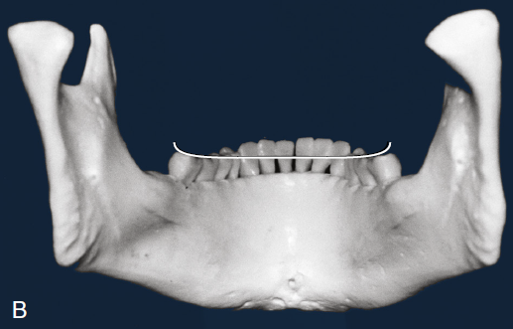
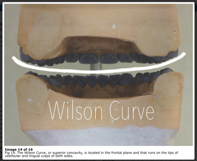
Curve of Wilson
The curve of Wilson is the cross-arch curvature of the posterior occlusal plane. The downward curvature of the arc is defined by a line drawn across the occlusal surface of the left mandibular first molar extending across the arch through the occlusal surface of the right mandibular first molar.
 |
 |
| 3D scan of a lower jaw model showing the dental arch from a frontal perspective with a wire illustrating the Curve of Wilson, which depicts the curvature across the tips of the molars. | A dental model illustrating the Wilson Curve with a line across the occlusal surfaces of both the upper and lower dental arches, highlighting the superior concavity in the frontal plane. |
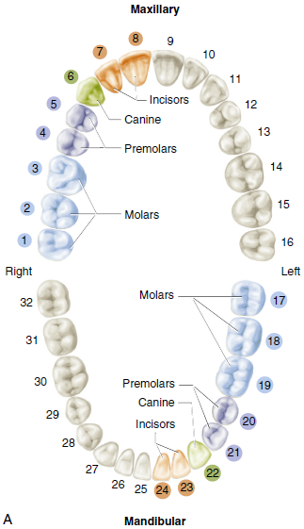
Tooth-Numbering Systems
Numbering systems are used as a simplified means of identifying the teeth for charting and descriptive purposes.
There are three basic systems:
- Universal/National System
- Palmer Notation System
- International Standards Organization System (ISO)
The system used most often in Canada is the International Standards Organization. This is the system that is used throughout this course. When charting and applying a tooth numbering system, we always start in the maxillary right, then move to the maxillary left, down to the mandibular left, and around to the mandibular right. This goes for permanent and primary dentition.
Universal/National System
This is the system that is used most often in the United States. Permanent teeth are numbered from 1 to 32. Numbering begins with the upper-right third molar and works around to the upper-left third molar. Then, numbering drops to the lower-left third molar and works around to the lower-right third molar.
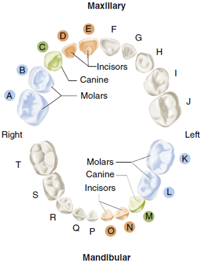
The primary dentition is lettered with upper case letters from A to T. Lettering begins with the upper-right primary second molar and works around to the upper-left primary second molar. Then, it continues by dropping to the lower-left primary second molar and works around to the lower-right primary second molar.
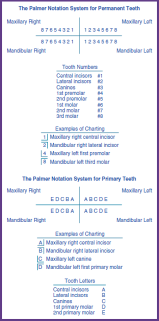
Palmer Notation System
A bracket designates one of the four quadrants in the Palmer Notation System. The bracket comprises a horizontal line separating the maxillary and mandibular arches and a vertical line representing the imaginary midline that runs between the central incisors and divides the oral cavity into left and right. The brackets are shown in red in the diagram to the right.
The brackets are to represent the quadrant that the tooth is in. The permanent teeth are numbered from 1 to 8 with 1 being the central incisors and 8 being the third molar. For the primary teeth are designated with a letter with A being the central incisor and E being the second molar.
International Standards Organization System
The ISO System uses a two-digit tooth-recording system. The first digit indicates the quadrant, and the second digit indicates the tooth within the quadrant, numbering from the midline toward the posterior.
In permanent dentition, the:
- Maxillary right quadrant is 1
- Maxillary left quadrant is 2
- Mandibular left quadrant is 3
- Mandibular right quadrant is 4
The teeth for all quadrants are number 1 to 8, with 1 being the central incisors working towards the back and ending with 8 representing the third molars.
Example: maxillary right central incisor is tooth 11, and mandibular left second premolar is tooth 35.
In the primary dentition, the:
- Maxillary right quadrant is 5
- Maxillary left quadrant is 6
- Mandibular left quadrant is 7
- Mandibular right quadrant is 8
The teeth for all quadrants are number 1 to 5, with 1 being the central incisors working towards the back and ending with 8 representing the second molars.
Example: maxillary right central incisor is tooth 51, and mandibular left second molar is tooth 75.
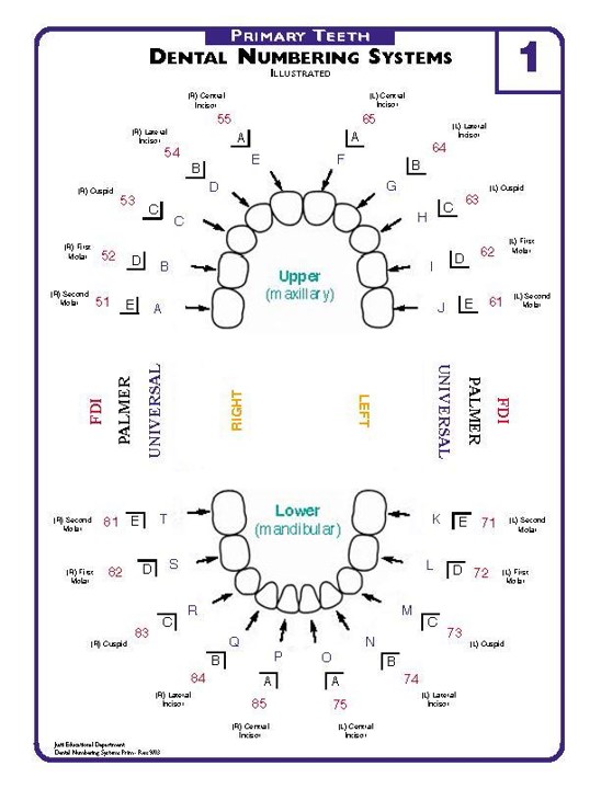 |
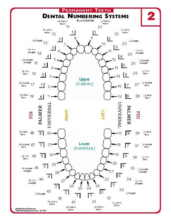 |
| Numbering systems for primary teeth. | Numbering systems for permanent teeth. |
You have completed Module 7A. Please return to Blackboard for the next steps.
Media Attributions
- Images from: Modern Dental Assisting, 13th and 14th Edition
(oe-KLOO-zhun); Natural contact of the maxillary and mandibular teeth in all positions.
(MAL-o-kloo-zhun); Occlusion that is deviated from a class I normal occlusion.
Maximum contact between the occluding surfaces of the maxillary and mandibular teeth.
Contact of the teeth during biting and chewing movements.
System developed by Dr. Edward H. Angle to describe and classify occlusion and malocclusion.
Ideal mesiodistal relationship between the jaws and the dental arches.
(DIS-toe-kloo-shun); Class II malocclusion in which the mesiobuccal cusp of the maxillary first molar occludes (by more than the width of a premolar) mesial to the mesiobuccal groove of the mandibular first molar.
Position in which the maxillary incisors are behind the mandibular incisors.
(MEE-zee-oe-kloo-zhun); Term used for class III malocclusion.
