Overview of Dentitions
Dentition Periods
People have two sets of teeth throughout their lifetime. You can observe substantial growth and change in the face and jaws as a person grows. The teeth also change in shape and size to accommodate this growth and development. The primary dentition is followed by the mixed dentition and then the permanent dentition. Although there are only two sets of teeth, there are three dentition periods:
| Dentition Period | Approximate Time Span | Teeth Marking Start of Period | Teeth Present in Oral Cavity |
| Primary | 6 months to 6 years | Eruption of first primary tooth | Primary teeth only |
| Mixed | 6 years to 12 years | Eruption of first permanent floor | Primary and permanent teeth |
| Permanent | 12 years and up | Exfoliation of last primary tooth | Permanent teeth only |
Primary Dentition
Let’s review some of the facts about primary dentition (the first set of 20 teeth to erupt in a child’s mouth):
- When others talk about the primary teeth, they may refer to them as “baby teeth,” “milk teeth,” or “deciduous teeth.”
- The primary dentition period lasts from approximately age 6 months through 6 years of age.
- The period begins when the lower central incisors erupt and ends when the first permanent molar erupts.
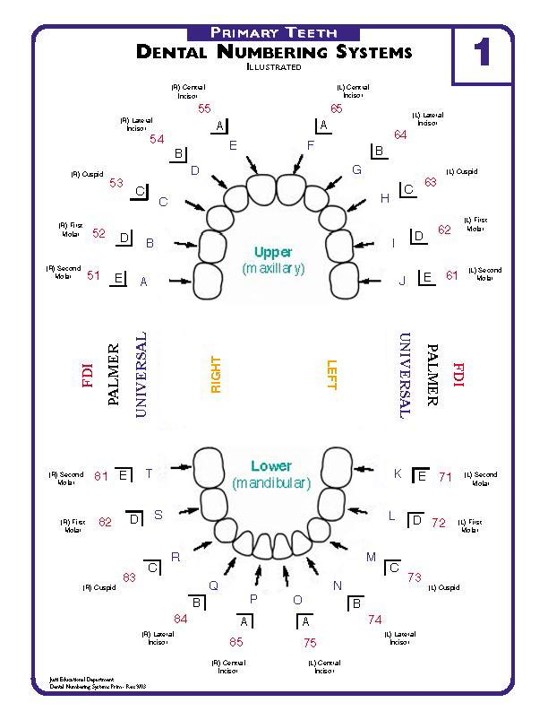 |
ERUPTION DATES
Primary Dentition – Dates for primary eruption are shown in months. Maxillary Teeth: Central Incisor 6-10 Lateral Incisor 9-12 First Molar 12-18 Canine 16-22 Second Molar 24-32 Mandibular Teeth: Central Incisor 6-10 Lateral Incisor 7-10 First Molar 12-18 Canine 16-22 Second Molar 20-32 |
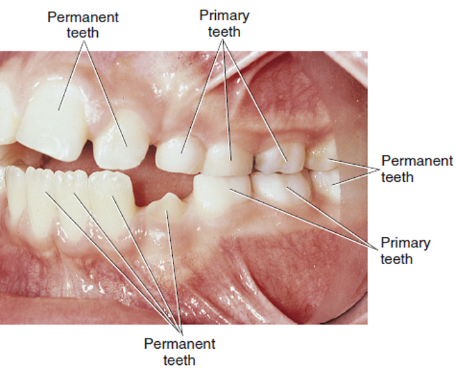
Mixed Dentition
Some of the facts about mixed dentition:
- Mixed dentition usually occurs between the ages of 6 and 12 years.
- Primary and permanent teeth are both present during this period.
- It begins with the eruption of the first permanent tooth and ends with the shedding of the last primary tooth.
- It may be a difficult time for children because of the colour (primary teeth are whiter than permanent teeth) and size differences between primary and permanent teeth.
Permanent Dentition
Now let’s review some of the facts about permanent or adult, dentition:
- The permanent dentition period begins at approximately 12 years of age after the last primary tooth has exfoliated.
- There are 32 teeth in the permanent dentition.
- Permanent incisors, canines, and premolars replace or “succeed” primary teeth and are called succedaneous teeth. Permanent molars are not succedaneous because they erupt behind (or posterior to) the primary second molars.
- After the eruption of the permanent canines and premolars and the second molars, the permanent dentition is complete at about age 14 years, except for the third molars, which do not erupt until age 18 to 25. Third molars do not always develop.
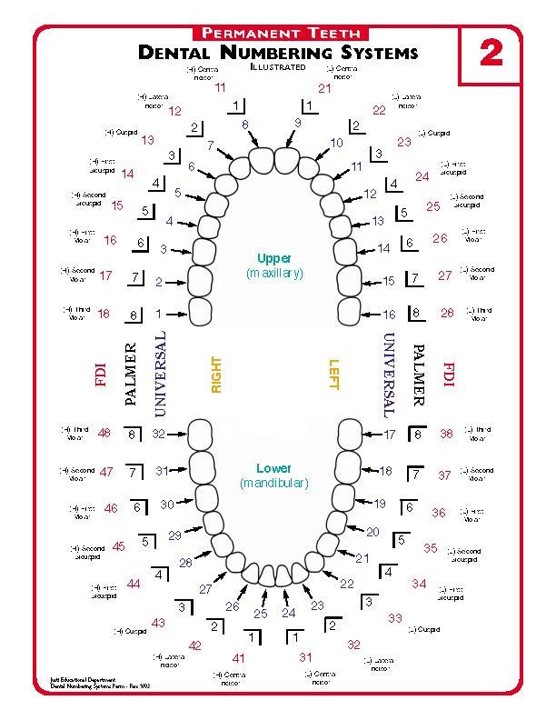 |
ERUPTION DATES
Permanent Dentition – Dates for permanent dentition shown in years. Maxillary Teeth: First molar 6-7 Central incisor 7-8 Lateral incisor 8-9 First premolar 10-11 Canine 11-12 Second premolar 12-13 Second molar 12-13 Third molar 17-21
Mandibular Teeth: First molar 6-7 Central incisor 6-7 Lateral incisor 7-8 Canine 9-10 First premolar 10-11 Second premolar 12-13 Second molar 11-13 Third molar 17-21 |
Dental Arches
The oral cavity has two arches: the maxillary arch and the mandibular arch. The maxillary arch is part of the skull, is fixed, and incapable of movement. The teeth are set in the maxilla bone. The maxillary arch may be referred to as the “upper arch.”
The mandibular arch is moveable through the action of the temporomandibular joint. The teeth are set in the mandible bone. The mandibular arch may be referred to as the “lower arch.” Occlusion is the contact between the maxillary and mandibular teeth.
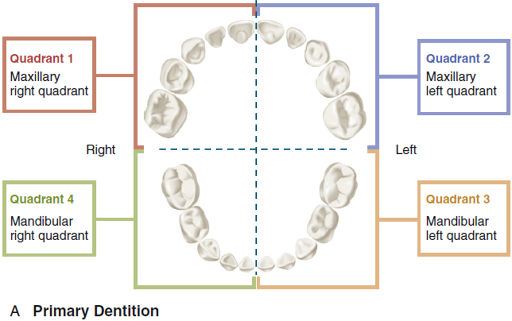
Quadrants
Dividing the maxillary and mandibular arches into halves yields four sections, which are called quadrants:
- Maxillary right quadrant
- Maxillary left quadrant
- Mandibular left quadrant
- Mandibular right quadrant
Each quadrant of permanent dentition contains eight permanent teeth, and each quadrant of primary dentition contains five teeth.
Remember, as you look into a patient’s oral cavity, the directions are reversed because you are facing each other. The patient’s left side is on your right, and the patient’s right side is on your left.
 |
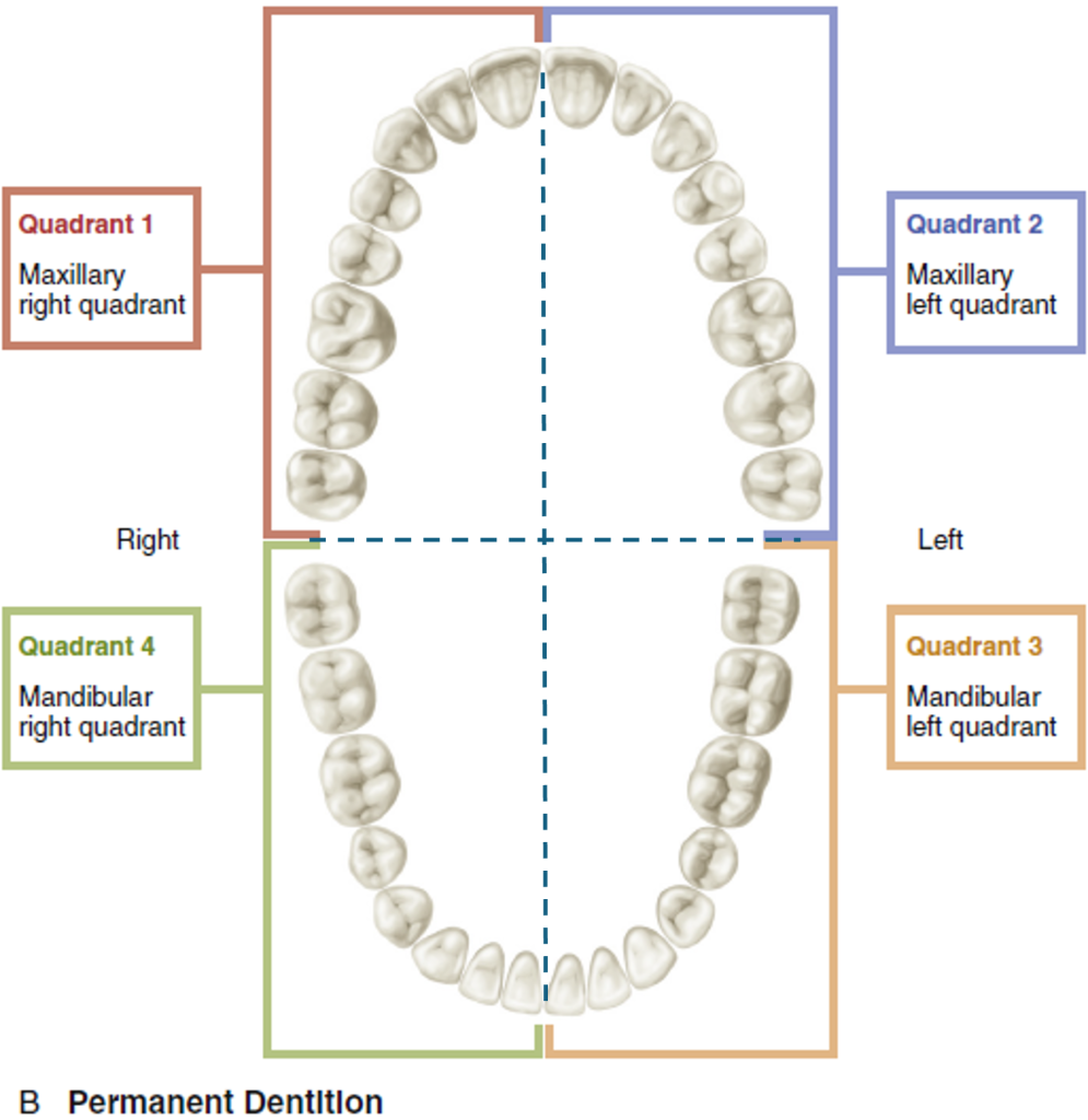 |
| Illustration showing an occlusal view of primary dentition divided into four quadrants: Quadrant 1, Maxillary right; Quadrant 2, Maxillary left; Quadrant 3, Mandibular left; and Quadrant 4, Mandibular right, with each quadrant color-coded and labeled. | Illustration showing an occlusal view of permanent dentition divided into four quadrants: Quadrant 1, Maxillary right; Quadrant 2, Maxillary left; Quadrant 3, Mandibular left; and Quadrant 4, Mandibular right, with each quadrant color-coded and labeled. |
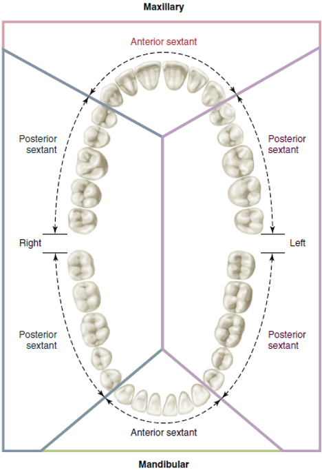
Sextants
Each arch can also be divided into sextants rather than quadrants. A sextant is one-sixth of the dentition. There are three sextants in each arch. The arches are divided as follows:
- Maxillary right posterior
- Maxillary anterior
- Maxillary left posterior
- Mandibular right posterior
- Mandibular anterior
- Mandibular left posterior
Anterior and Posterior Teeth
Another way to identify the location of teeth in the mouth is to describe them as anterior or posterior.
Anterior teeth (teeth toward the front of the mouth) include the central incisors, lateral incisors, and canines. The anterior teeth are aligned in a gentle curve.
Posterior teeth (teeth toward the back of the mouth) in the primary dentition include the molars. In the permanent dentition, the posterior teeth include premolars and all molars. The posterior teeth appear in an almost straight line (one behind the other).
Recalling how the teeth are aligned is important when exposing radiographs.
Types and Functions of Teeth
Human beings eat both meat and plants. To accommodate this variety in the diet, teeth are designed to cut, tear, and grind different types of food.
The permanent dentition is divided into four types of teeth:
- Incisors
- Canines
- Premolars
- Molars
The primary dentition has:
- Incisors
- Canines
- Molars
There are no premolars in the primary dentition.
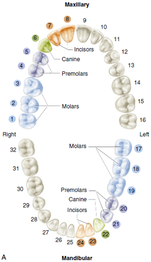
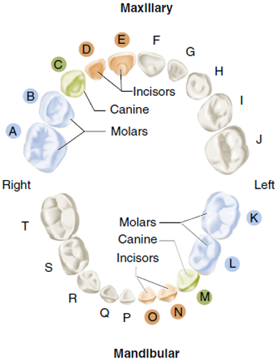
Occlusal Views of Permanent and Primary Dentition
The diagram on the right displays occlusal views of maxillary and mandibular arches with permanent teeth. The diagram on the left displays occlusal views of primary teeth in maxillary and mandibular arches.
 |
 |
Incisors
Incisors are single-rooted teeth with a relatively sharp, thin edge. Located at the front of the incisors, they are designed to cut food without applying heavy force. Incisor means “that which makes an incision or cut.” The incisors’ lingual surface (tongue side) is shaped like a shovel to help guide food into the mouth.
Canines
Canine teeth, also known as cuspids, are located at the “corner” of the arch. They are designed for cutting and tearing food, which requires the application of force.
Canines are the longest teeth in the human dentition. They have the longest roots of all teeth and are usually the last teeth to be lost. Because of its sturdy crown, long root, and location in the arch, the canine is referred to as the cornerstone of the dental arch.
Premolars
There are four maxillary and four mandibular premolars. The premolars, also known as bicuspids, are a cross between canines and molars. Bicuspid is inaccurate as it refers to two (bi) cusps; some premolars have three cusps. The pointed buccal cusps hold the food while the lingual cusps grind it. Premolars are not as long as canines and have a broader surface for chewing food. There are no premolars in the primary dentition.
Molars
Molars are much larger than premolars, usually having four or more cusps. The function of the 12 molars is to chew or grind food. Depending on the tooth’s location, there are four or five cusps on the occlusal (biting) surface of each molar.
Maxillary and mandibular molars differ significantly in shape, size, and number of cusps and roots.
Tooth Surfaces Relative to Other Oral Cavity Structures
As mentioned previously, you can identify most tooth surfaces by their relationship with other structures in the oral cavity. Let’s take a closer look at those relationships now.
The facial surface is the surface closest to the face. This surface may also be referred to as labial (closest to the lips) on anterior teeth or buccal (closest to the cheek) on posterior teeth.
On maxillary teeth, the lingual surface may be called the palatal surface. The incisal surface (incisal edge) is the biting surface of the anterior teeth. Teeth touch one another in the dental arch. These surfaces are referred to as proximal surfaces.
If a tooth’s proximal surface is near or toward the midline, it is called the mesial surface. If a tooth’s proximal surface is distant from the midline, it is called distal. Posterior teeth have occlusal surfaces that grind the food when chewing. The lingual surface is the surface closest to the tongue.
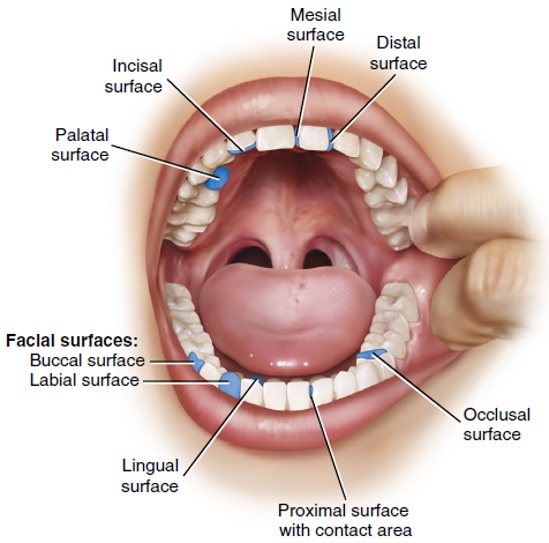
Tooth Surfaces
When trying to visualize the various surfaces of a tooth, it is helpful to imagine the tooth as a box with sides. Each tooth has five surfaces, most of which are identified by their relationship to other oral structures. The five tooth surfaces are:
| Anterior Teeth | Posterior Teeth | ||
| Surface Name | Meaning/Location | Surface Name | Meaning/Location |
| Facial or labial | Closest to face or lips | Facial or buccal | Closest to face or cheek |
| Lingual | Closest to the tongue | Lingual | Closest to the tongue |
| Incisal | Surface used to bite | Occlusal | Surface used to chew |
| Mesial | Closest to the midline | Mesial | Closest to the midline |
| Distal | Distant from the midline | Distal | Distant from the midline |
Activity 1: Tooth Surfaces
Anatomical Features of Teeth
All teeth have contours, contacts, and embrasures. These features help maintain the teeth in their position in the arch and protect the tissues during mastication (chewing).
Test Your Knowledge
Contours
All teeth have a curved surface except when the tooth is fractured or worn. Some surfaces are convex; others are concave. Contours may vary; the general principle that the tooth’s crown narrows toward the cervical line is valid for all types of teeth.
Facial and Lingual Contours
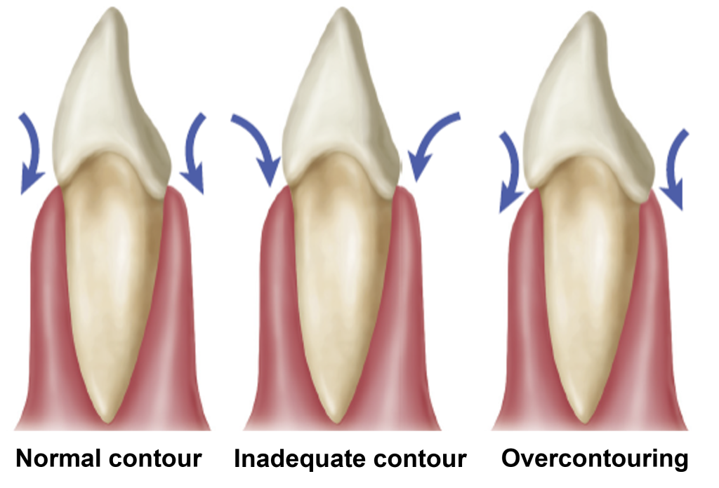
Curvatures on the facial and lingual surfaces provide natural passageways for food. This action protects the gingiva from the impact of food during chewing. The normal contour of a tooth provides the gingiva with adequate stimulation for health while protecting it from damage that may be caused by food.
When a tooth is restored, it is important to contour the tooth so that it is the same as it previously was. When a tooth has inadequate contours, the gingiva may be traumatized by food pushing against it. If a tooth is over-contoured, the gingiva will lack stimulation and may be challenging to keep clean.
Mesial Distal Contours
Contours of the mesial and distal surfaces provide regular contact and embrasure form. These contours are considered self-cleansing and contribute to the self-preservation of the tooth.
Tooth Contours
The diagram below shows tooth contours, which are a normal contour, an inadequate contour, and an overcontouring. Every tooth has a curved surface except when the tooth is fractured or worn. Some surfaces are convex (curved outward); others are concave (curved inward). Although the general contours vary, the general principle that the tooth’s crown narrows toward the cervical line is true for all types of teeth.
Contacts
The contact area is the area of a tooth’s mesial or distal surface that touches the adjacent tooth in the same arch. The contact point is the exact spot at which teeth each other. Contact and contact area may be used interchangeably to refer to the contact point.
The crown of each tooth in the maxillary and mandibular arches should be in contact with the adjacent tooth or teeth. A proper contact serves the following purposes:
- Prevents food from being trapped between the teeth.
- Stabilizes the dental arches by holding the teeth in either arch in positive contact with each other.
- Protects the interproximal gingival tissue from trauma during mastication.
When adjacent teeth do not touch one another, it is called open contact.
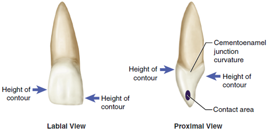
Height of Contour
The height of the contour is the “bulge,” or widest point, on a specific crown surface. Contact areas on the mesial and distal surfaces are usually considered the height of contour on the proximal surfaces. Facial and lingual surfaces also have a height of contour.
When a removable partial denture is fabricated, it utilizes these contour heights for retention. The clasps stretch to fit over these contour heights and hold the removable partial denture in place.
The diagram below shows an example of a permanent anterior tooth with the contact area and the height of the contour identified. Note that the height of the contour is not the same on this tooth’s buccal and lingual surfaces.
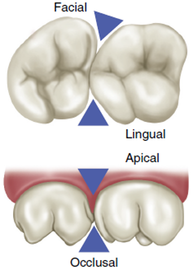
Embrasures
An embrasure is a triangular space in a gingival direction between the proximal surfaces of two adjoining teeth in contact. Embrasures are continuous with the interproximal spaces between the teeth. Embrasures may diverge facially, lingually, occlusally, or apically.
All tooth contours, including contact areas and embrasures, are important in the function and health of the oral tissues.
Reflect: Can you feel the embrasures of your teeth with your tongue?
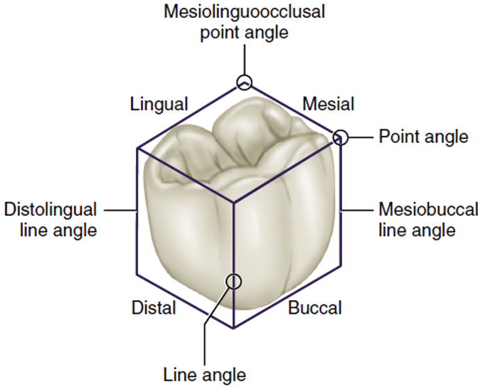
Angles and Divisions of Teeth
The crowns and roots are divided into thirds for a more precise description of teeth. The junctions of the crown surfaces are described as line angles and point angles.
A line angle is formed by the junction of two surfaces and is named from the combination of the two joined surfaces. Example: On an anterior tooth, the junction of the mesial and labial surfaces is called the mesiolabial line angle.
A point angle is formed by the junction of three surfaces at one point and is Named from the combination of surfaces that form them. Example: The junction of the mesial, buccal, and occlusal surface of a molar is called the mesiobucco-occlusal point angle.
Line angles and point angles are used only as descriptive terms to indicate specific locations. When combining the names of the surfaces, the last two letters of the first and/or second surface are dropped, and the letter ‘o’ is substituted.
Line and Point Angles
The diagram below shows line and point angles. For a better description of the teeth, the crowns and roots of the teeth have been divided into thirds, and junctions of the crown surfaces are described as line angles and point angles.
Divisions into Thirds
Each tooth surface is divided into imaginary thirds to help identify a specific area on a tooth. These thirds are named according to the areas they approximate. The diagram below shows an anterior tooth and a posterior tooth, with designations for crown and root thirds.
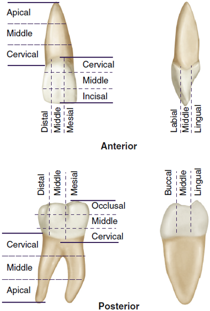
The tooth’s root is divided crosswise into thirds: the apical third (nearest the tip of the root), middle third, and cervical third (nearest the neck of the tooth).
The crown of a tooth is divided into thirds in three divisions:
- Occlusocervical division: The crosswise division parallel to occlusal or incisal surface. The occlusocervical division consists of the occlusal third, middle third, and cervical third.
- Mesiodistal division: The lengthwise division in a mesial-distal (front to back) direction. The mesiodistal division consists of the mesial third, middle third, and distal third.
- Buccolingual division: The lengthwise division in a labial or buccal-lingual direction. The buccolingual division consists of the facial or buccal/labial third, middle third, and lingual third.
You have completed Module 7. Please return to Blackboard for the next steps.
Media Attributions
- Images from: Modern Dental Assisting, 13th and 14th Edition
First set of 20 primary teeth.
Mixture of permanent teeth and primary teeth that occurs until all primary teeth have been lost, usually between ages 6 and 12.
Set of 32 secondary teeth.
(duh-SID-yoo-us); pertaining to first dentition of 20 teeth; often called “baby teeth” or primary teeth.
(suk-se-DAY-nee-us); Permanent teeth that replace primary teeth.
Toward the back.
(MAK-si-lar-ee); Upper jaw.
(man-DIB-you-ler); Lower jaw.
(oe-KLOO-zhun); Natural contact of the maxillary and mandibular teeth in all positions.
One quarter of the dentition.
One sixth of the dentition.
Toward the front.
Surface of mandibular and maxillary teeth closest to the tongue; also called palatal surface.
Region of the head that refers to structures closest to the inner cheek.
Chewing surface of posterior teeth.
Tooth surface closest to the face. Facial surfaces closest to the lips are called labial surfaces, and facial surfaces closest to the inner cheek are called buccal surfaces; therefore the term facial can be substituted for labial and buccal, and vice versa.
Lingual surface of maxillary teeth.
Chewing surface of anterior teeth.
Surfaces next to each other when teeth are adjacent in the arch.
Surface of the tooth toward the midline.
Farther away from the trunk of the body; opposite of proximal.
Triangular space in a gingival direction between the proximal surfaces of two adjoining teeth in contact.
Curved outward.
Curved inward.
Area of the mesial or distal surface of a tooth that touches the adjacent tooth in the same arch.
(in-tur-PROK-si-mul); Space area between adjacent tooth surfaces.
Junction of two tooth surface walls.
Angle formed by the junction of three surfaces.
Division of the root nearest the tip of the root.
Of the root in the middle.
Division of the root nearest the neck of the tooth.
Crosswise division of the crown that is parallel to the occlusal or incisal surface, consisting of the occlusal third, middle third, and cervical third.
Lengthwise division of the crown in a mesiodistal (front-to-back) direction, consisting of the mesial third, middle third, and distal third.
Division lengthwise division of the crown in a labial or buccolingual direction, consisting of the facial or buccal/labial third, middle third, and lingual third.
