Landmarks of the Face and Oral Cavity
A dental assistant must be thoroughly knowledgeable about the landmarks of the face and oral cavity. In addition to serving as helpful reference points for dental radiography and other procedures, facial features provide essential landmarks for many deeper structures. Any deviation from normal surface features may be clinically significant. You may wish to examine your own face and mouth to assist you in identifying some of these landmarks.
Landmarks of the Face
The face is defined as the part of the head visible in a frontal view and anterior to the ears, and all that lies between the hairline and the chin.
The face can be subdivided into nine areas, which are:
- Forehead: Extending from the eyebrows to the hairline
- Temples: Lateral to the eyes
- Orbital: Eye area that is covered by the eyelids
- External nose
- Zygomatic (malar): Prominence of the cheek
- Mouth and lips
- Cheeks
- Chin
- External ear
See the hotspot image below to see where the nine landmarks belong on the face.
Features of the Face
A dental assistant should be able to identify the following 13 important features of the face:
- Outer Canthus
- Inner Canthus
- Ala
- Philtrum
- Tragus
- Nasion
- Glabella
- Root
- Septum
- Anterior Naris
- Mental Protuberance
- Angle of the Mandible
- Zygomatic Arch
See the hotspot image below to see where the thirteen features belong on the face and learn a bit more about each feature is.
Skin
The skin of the face is thin to medium in relative thickness. It is soft and movable over a layer of loose connective tissue. The skin around the external ear and the ala of the nose is fixed to underlying cartilage. Facial skin contains many sweat and sebaceous glands. Below the skin is a layer of connective tissue that contains variable amounts of fat. This fat smooths out the face’s contours, particularly between facial expression muscles. Located within the connective tissue are the motor and sensory nerves of facial expression.
Lips
The lips, also called labia, provide the gateway to the oral cavity. The lips are formed externally by the skin and internally by the mucous membrane. The lips are outlined by the vermilion border, which is darker in colour than the surrounding skin. The vermillion border is the area where one would apply lip liner.
The labial commissure is the angle at the corner of the mouth where the upper and lower lips join.
The nasolabial sulcus is the groove extending upward between each labial commissure and the ala of the nose.
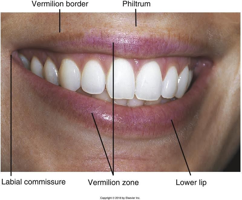
The normal appearance of the lips is seeing a defined border between the lips and the surrounding skin of the face (vermillion border). One will look for any loss of the vermillion border when examining the lips. If noted, it could be the result of scar tissue from a past injury, the result of sun exposure, or a sign of oral cancer. A biopsy would be required to diagnose cells.
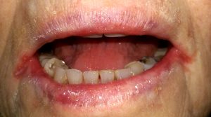
The image below is of a condition called angular cheilosis, which is an inflammation or cracking at the corners of the mouth. It is associated with a vitamin B deficiency.
Test Your Knowledge
Activity 1: Features of the Face
The Oral Cavity
The entire oral cavity is lined with mucous membrane tissue. This type of tissue is moist and is adapted to meet the needs of the area it covers.
The oral cavity proper consists of the following two areas:
- The vestibule is the space between the teeth and the inner mucosal lining of the lips and cheeks.
- The oral cavity proper is the space contained within the upper and lower dental arches.
The Vestibule
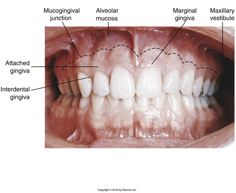
The vestibule begins on the inside of the lips and extends from the lips onto the alveolar process of both arches. The vestibules are lined with mucosal tissue. The vestibular mucosa is thin, red, and loosely bound to the underlying alveolar bone. The base of each vestibule, where the buccal mucosa meets the alveolar mucosa, is called the mucobuccal fold.
The mucogingival junction is a distinct line of color change where the alveolar membrane meets with the attached gingiva. The attached gingiva is lighter in colour and has a stippled appearance.
Tissues of the Oral Cavity
The inside surfaces of the cheeks form the side walls of the oral cavity. The area between the cheeks and the teeth or alveolar ridge is called the buccal vestibule. The parotid papilla is a small, elevation of tissue that is located on the inner surface of the cheek on the buccal mucosa. The parotid papilla protects the opening of the parotid duct (also called Stenson’s duct) of the parotid salivary gland.
 |
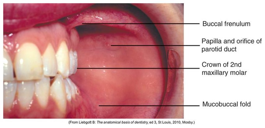 |
| Close-up image of an open mouth showing a detailed view of the oral cavity tissues, including the vestibular fold, labial frenulum, labial mucosa, alveolar mucosa, and labial gingiva for both maxillary and mandibular regions. | Close-up image of the inside of a mouth showing the buccal region with labeled anatomical features, including the buccal frenulum, papilla orifice of the parotid duct, crown of the second maxillary molar, and the mucobuccal fold. |
Normal Variations of the Buccal Mucosa
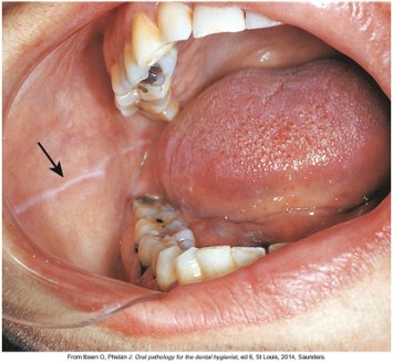
Fordyce’s spots are normal, small, yellowish elevations that may be seen on the buccal mucosa. Another normal variation that may be noted on the buccal mucosa is linea alba, a white ridge of raised tissue running horizontally in the area where the maxillary and mandibular teeth come together.
Labial and Lingual Frenum
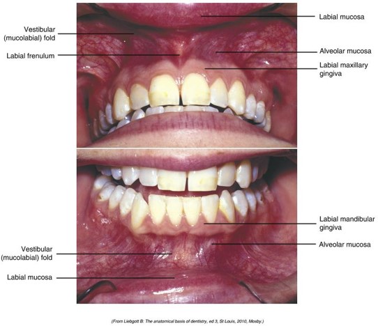
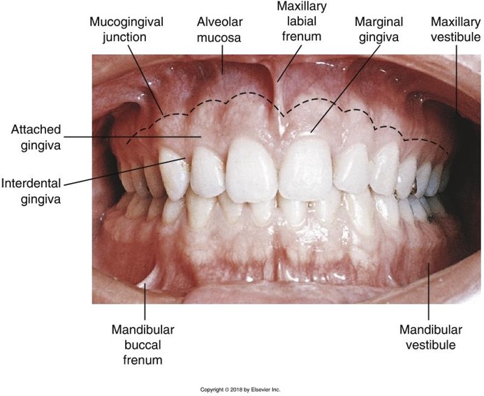
A frenum (plural is frenula) is a narrow band of tissue that connects two structures. The maxillary labial frenum passes from the midline of the maxillary arch to the midline of the inner surface of the upper lip. The mandibular labial frenum passes from the midline of the mandibular arch to the midline of the inner surface of the lower lip. The buccal frenum passes from the oral mucosa near the maxillary or mandibular first molars to the inner surface of the cheek. The lingual frenum passes from the floor of the mouth to the midline of the ventral (underside) of the tongue.
Below is an image of the fenula, displaying healthy teeth and gums identifying various parts such as the mucogingival junction, alveolar mucosa, maxillary and mandibular labial frenum, attached and interdental gingiva, and the maxillary and mandibular vestibules.
Gingiva
The gingiva (plural, gingivae) commonly referred to as the gums, are masticatory mucosa that cover the alveolar processes of the jaws and surround the necks of the teeth.
Normal gingival tissue has the following characteristics:
- It surrounds the tooth like a collar and is self-cleansing.
- It is firm, resistant, and can be tightly adapted to the tooth and bone.
- The surfaces of the attached gingivae and interdental papillae are stippled and similar in appearance to the rind of an orange.
- The surface color varies according to the individual’s pigmentation.
Sometimes, the gingivae do not completely cover the roots of the teeth, and the roots become exposed. This condition, called gingival recession, can lead to sensitivity and root cavities (caries).
 |
 |
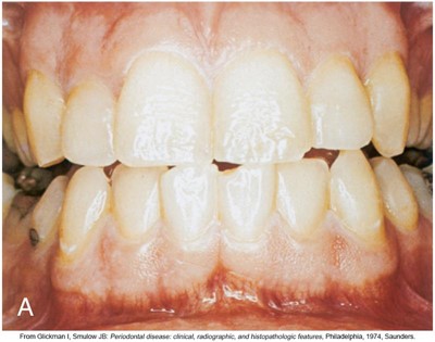
| Normal gingiva on a light-skinned individual. | Normal gingiva on a dark-skinned individual. |
Unattached Gingiva
Unattached gingiva, also known as marginal gingiva or free gingiva, is the border of the gingiva that surrounds the teeth in a collar-like fashion.
Unattached gingiva is:
- usually light pink or coral coloured
- is not bound to the underlying tissue of the tooth
- consists of tissues from the top of the gingival margin to the base of the gingival sulcus.
- usually about 1 mm wide and forms the soft wall of the gingival sulcus
- is the first tissue to respond to inflammation
Other Parts of the Gingiva
- Interdental Gingiva (also called gingival papilla): Extension of the free gingiva that fills the interproximal embrasure between two adjacent teeth
- Gingival Groove: Shallow groove that runs parallel to the margin of the unattached gingiva and marks the beginning of the attached gingiva
- Attached Gingiva: Extends from the base of the sulcus to the mucogingival junction
Reflect: With your tongue, can you feel the firmness of the attached gingiva compared with the softer alveolar mucosa of the vestibules?
Test Your Knowledge
Activity 2: Landmarks of the Face and Oral Cavity
You have completed Module 5. Please return to Blackboard for the next steps.
Media Attributions
- Images from: Modern Dental Assisting, 13th and 14th Edition
Fold of tissue at the corner of the eyelids.
Winglike tip of the outer side of each nostril; plural, alae.
Rectangular area from under the nose to the midline of the upper lip.
Cartilaginous projection anterior to the external opening of the ear.
Midpoint between the eyes just below the eyebrows.
Smooth surface of the frontal bone; also, the anatomical part directly above the root of the nose.
Facial landmark commonly called the “bridge” of the nose.
Tissue that divides the nasal cavity into two nasal fossae.
Nostril; plural, nares
Part of the mandible that forms the chin.
Lower posterior of the ramus.
Arch formed when the temporal process of the zygomatic bone articulates with the zygomatic process of the temporal bone.
Gateway to the oral cavity; commonly known as “lips”.
Darker-colored border around the lips.
The angle at the corner of the mouth where the upper and lower lips join.
Groove extending upward between the labial commissure and the nasal ala.
Space on the tongue side within the upper and lower dental arches.
Space between the teeth and the inner mucosal lining of the lips and cheeks.
Base of the vestibule where the buccal mucosa meets the alveolar mucosa.
Distinct line of color change in the tissue where the alveolar membrane meets with attached gingivae.
Masticatory mucosa that covers the alveolar processes of the jaws and surrounds the necks of the teeth; plural, gingivae.
Area between the cheeks and the teeth or alveolar ridge.
Small elevation of tissue located on the inner surface of the cheek.
Normal variations that may appear on the buccal mucosa
Normal variation noted on the buccal mucosa.
A narrow band of tissue that connects two structures (plural, frenulae).
Band of tissue that passes from the facial oral mucosa at the midline of the arch to the midline of the inner surface of the lip; also called frenulum; plural, frenula.
Thin fold of mucous membrane that extends from the floor of the mouth to the underside of the tongue.
