Tooth Morphology – Permanent Posterior Teeth (Part A)
An Overview of Posterior Permanent Dentition
The permanent posterior dentition includes premolars and molars. These teeth enable us to chew our food properly. Generally speaking, their crown and root anatomy is a great example of “form enables function.”
- Posterior teeth have large crowns and sturdy roots.
- Posterior teeth have large occlusal surfaces.
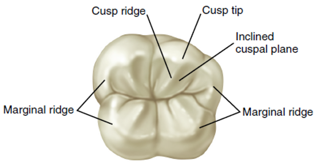
- Occlusal surfaces have anywhere from two to five cusps as needed for grinding. Imagine each cusp as a mountain with sloping areas, or cusp ridges, extending from the top of the mountain; sloping areas called inclined cuspal planes are between the ridges.
- Occlusal surfaces have multiple fossae, pits, and grooves to hold the food on the occlusal table during chewing.
Unfortunately, these pits and grooves are highly susceptible to decay if they are not brushed correctly and regularly. It is common for children between the ages of 6 and 14 to have sealants applied to the posterior teeth to prevent tooth decay.
Each shallow, wide depression on the occlusal table is a fossa. One type of fossa on posterior teeth, the central fossa, is located where the cusp ridges converge in a central point, where the grooves meet. Another type of fossa is the triangular fossa. Sometimes located in the deepest portions of the fossa are occlusal developmental pits. Each pit is a sharp pinpoint depression where two or more grooves meet.
Occlusal Surface of a Permanent Posterior Tooth
 |
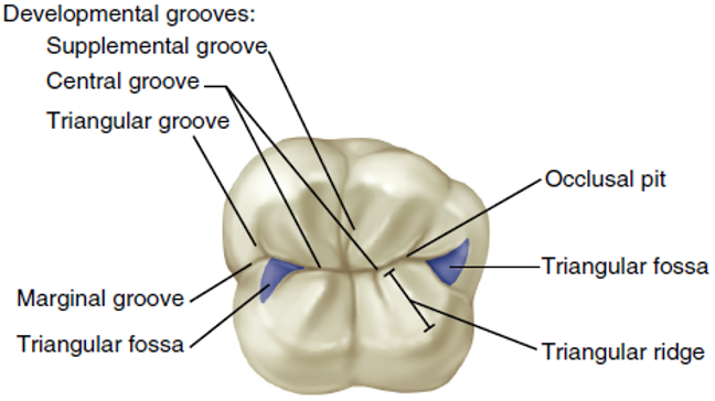 |
| A diagram of a permanent posterior tooth’s occlusal surface highlights anatomical features such as the cusp tip, cusp ridge, inclined cuspal plane, and marginal ridges. | A diagram of a molar tooth occlusal surface indicating various anatomical structures, including developmental grooves (supplemental, central, triangular), occlusal pit, marginal groove, triangular fossa, and triangular ridge. |
Maxillary First Premolars – Teeth 14 and 24
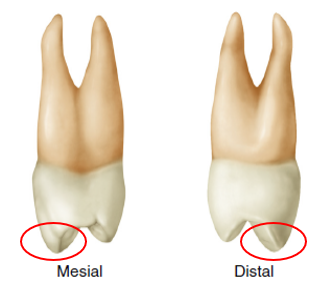
Maxillary first premolars are larger than maxillary second premolars. Each maxillary first premolar has two cusps (buccal and lingual) and a bifurcated root (facial and lingual). Both maxillary premolars erupt earlier than the mandibular premolars.
If you look at the maxillary first premolar from the occlusal view, you will see that the cusps are separated by a prominent central developmental groove (indicated by the blue line). The mesial groove from the central developmental groove crosses the mesial marginal ridge onto the mesial surface to form the anatomical landmark called the mesiomarginal developmental groove. This will help you distinguish between and right and left first premolars. Permanent maxillary first premolars are the succedaneous replacements for the primary maxillary first molars.
 |
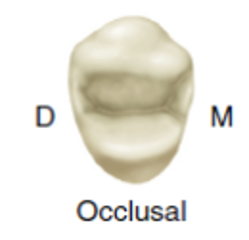 |
| Maxillary first premolars have two distinguishing features. These teeth have two cusps (buccal and lingual) and a bifurcated root. Note that the buccal cusp is longer and larger than the lingual cusp. | The occlusal table is defined by the four cusp ridges, a mesial marginal ridge, and a distal marginal ridge. |
Maxillary Second Premolars – Teeth 15 and 25
It is easy to see the differences when comparing the maxillary second premolar to the maxillary first premolar. Permanent maxillary second premolars are the succedaneous replacements for the primary maxillary second molars.
 |
 |
 |
| The maxillary second premolar has buccal and lingual cusps that are more equal in size and length. | The maxillary second premolar usually has only one root. | The occlusal surface of the maxillary second premolar has numerous supplemental grooves rather than one prominent central developmental groove. Where the first premolar has a mesial and distal fossa on the occlusal surface, the second premolar has mesial and distal ridges. |
Mandibular First Premolar – Teeth 34 and 44
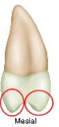
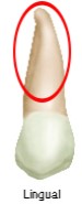
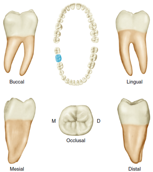
The mandibular first premolar can sometimes be mistaken for a mandibular canine. This is because it has a large, well-developed buccal cusp and a small, nonfunctional lingual cusp that looks like a large cingulum. These two characteristics combine to give the appearance of a very small occlusal table. The mandibular first premolar has only one root.
Permanent mandibular first premolars are the succedaneous replacements for the primary mandibular first molars.
 |
 |
| The mandibular first premolar has a large, well-developed buccal cusp and a small, nonfunctional lingual cusp resembling a large cingulum. | The mesial section of the central developmental groove extends onto the lingual surface, forming the mesiolingual developmental groove. This will help you distinguish between right and left first premolars. |
Mandibular Second Premolar – Teeth 35 and 45
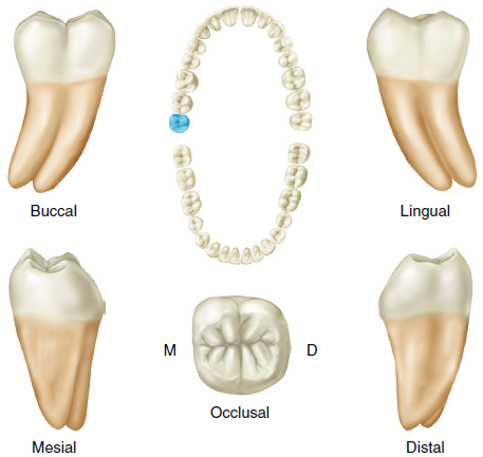
Mandibular second premolars have the largest occlusal surfaces of all the premolar teeth. This tooth may have two cusps (bicanineate) or three cusps (tricanineate) with different groove patterns.
Groove patterns on the occlusal surface of a two-cusp mandibular second premolar are in the shape of either the letter U or the letter H, and the groove pattern on the three-cusp tooth (the most common) is in the shape of the letter Y. Permanent mandibular second premolars are the succedaneous replacements for the primary mandibular second molars.
See the hotspot image below for different occlusal views of a permanent mandibular second premolar. Note the single deep groove in the two-cusp teeth compared with the distinctive Y pattern of the three-cusp tooth.
Activity 1: Characteristics of Permanent Premolars
Overview of Maxillary Molars
Maxillary molars have several common characteristics:
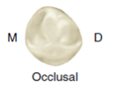
- Maxillary molars have a rhomboid (twisted square-shaped) occlusal surface that is widest in the buccal-lingual dimension.
- Each maxillary molar usually has four major cusps, with two on the buccal portion of the occlusal table and two on the lingual.
- Maxillary molars have trifurcated (three) roots, one on the lingual and two on the buccal.
- The roots of the maxillary molars may penetrate the maxillary sinus as a result of accidental trauma or during an extraction.
- The permanent maxillary third molars may fail to erupt and may remain impacted within the alveolar bone.
- If the maxillary first molar is lost, the second molar can tip and drift into the open space, causing difficulty in chewing and furthering periodontal disease.
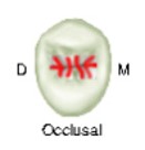
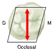
Below is a diagram of an occlusal view of a maxillary molar with a diagrammatic hexagonal outline and a double-headed red arrow indicating the tooth’s mesiodistal dimension, labeled ‘M’ for mesial and ‘D’ for distal.
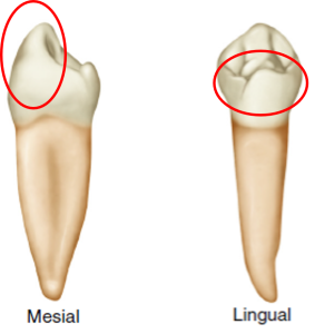
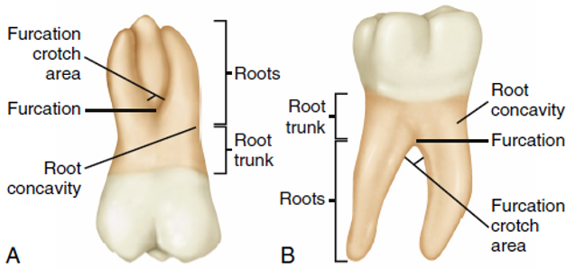
Below is a diagram of A: maxillary first molar and B: mandibular first molar. The three roots on the maxillary molar are compared with two on the mandibular, and the mandibular molars tend to be wider than the maxillary ones, with larger cusps.
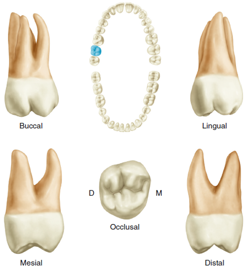
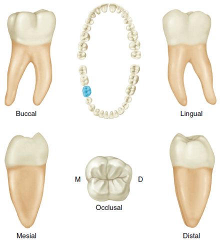
Views of a Permanent Maxillary Right First Molar
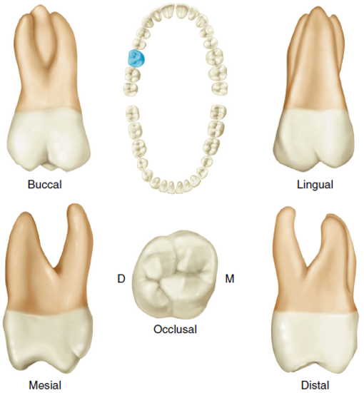
Below is a diagram that displays multiple views of a permanent maxillary right first molar, including buccal, lingual, mesial, distal, and occlusal. A dental arch is also shown, highlighting the position of the molar.
Maxillary First Molars – Teeth 16 and 26
The first molars of the maxillary are the first permanent teeth to erupt into the maxillary arch. Maxillary first molars erupt distal to the primary maxillary second molars and are, therefore, nonsuccedaneous (do not replace the primary teeth).
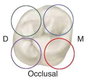
The first maxillary molar is the largest tooth in the maxillary arch and has the largest crown in permanent dentition. It comprises four well-developed cusps and one supplemental fifth cusp called the cusp of carabelli.
A distinguishing feature of maxillary molars is an oblique angle pattern on the occlusals. It extends from the distobuccal to the mesiolingual and includes a ridge, groove, and fossa. This landmark will help you determine right and left maxillary molars.
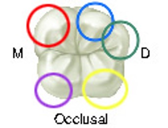
The first molar of the maxillary is the largest tooth in the maxillary arch. The tooth in the diagram below has four well-developed functional cusps (listed from largest to smallest):
- Mesiolingual (red circle on the image)
- Mesiobuccal (blue circle on the image)
- Distobuccal (green circle on the image)
- Distolingual (purple circle on the image)
Maxillary Second Molars – Teeth 17 and 27
The maxillary second molars supplement the first molars in function. The maxillary second molars erupt distal to the maxillary first molars and are nonsuccedaneous. The crown of the maxillary second molar has four cusps and is slightly shorter than the maxillary first molar. There is no fifth cusp present. The mesiobuccal cusp is slightly longer than the distobuccal cusp. The roots of the maxillary second molars are smaller than the roots of the maxillary first molars. The lingual root is the largest and longest.
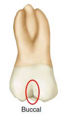
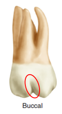
The buccal groove is located farther distally on the buccal surface of the second maxillary molar than on the first maxillary molar.
 |
 |
| Max. 1st Molar | Max. 2nd Molar |
The maxillary second molar has three roots:
|
 |
The maxillary second molar has four cusps (listed largest to smallest):
|
Maxillary Third Molars – Teeth 18 and 28
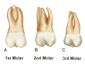
Maxillary third molars often appear as an anomaly. The third molars differ considerably in size, contour, and relative position from the other teeth. Maxillary third molars are more likely to be out of position in the arch compared to other teeth. The crown of the maxillary third molar is smaller, and the roots are usually shorter than those of the other maxillary molars. The roots of the maxillary third molar tend to fuse, resulting in a single tapered root.
People sometimes refer to the maxillary third molars as the “wisdom” teeth because they erupt last. These teeth are the most likely to be missing due to heredity or congenital causes.
The image below shows buccal views of permanent maxillary right molars. Notice how the roots are closer when the molars are farther apart. Third molar roots are often fused. Cusp size and root length get smaller/shorter from the first molar to the third molar. The first molar is the largest and longest, and the third molar is the smallest and shortest of the three molars.
Mandibular First Molars – Teeth 36 and 46
The first molars of the permanent mandibular are usually the first permanent teeth to erupt into the oral cavity. They erupt distal to the primary second molars and are, therefore, nonsuccedaneous. The mesiobuccal cusp is the largest, widest, and highest cusp on the buccal side. The distobuccal cusp is slightly smaller, shorter, and sharper. The distal cusp is the lowest cusp. The mandibular first molars have two roots, mesial and distal. The roots of a mandibular first molar are larger and more divergent than those of a second molar.
The permanent mandibular first molars have five well-developed cusps that are circled in the tooth diagram below:
- Mesiobuccal (red circle in the image)
- Mesiolingual (purple circle in the image)
- Distolingual (yellow circle in the image)
- Distobuccal (blue circle in the image)
- Distal (green circle in the image)
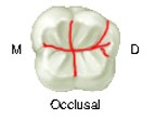
Developmental grooves separate the five cusps, as shown by the red lines on the occlusal surface image below.
Mandibular Second Molars – Teeth 37 and 47
The mandibular second molars erupt between 11 and 12 years of age. These teeth erupt distal to the permanent first molars and are nonsuccedaneous.
The crown of the mandibular second molar is slightly smaller than that of the first molar in all directions. The crown has four well-developed cusps (mesiolingual, distolingual, mesiobuccal, and distobuccal). All four cusps are usually the same shape and size. There is no distal cusp on the mandibular second molars. The mandibular second molars have two roots. The roots are slightly shorter and less divergent than the roots of the mandibular first molars.
Mandibular Third Molars – Teeth 38 and 48
The mandibular third molars are similar to the maxillary third molars in that they vary greatly in shape. There is no typical mandibular third molar. This molar is usually smaller in all dimensions than the second molar.
The crown of the mandibular third molars consists of four developmental lobes taper distally when viewed from the mesial aspect. The two mesial cusps are larger than the distal cusps, and the occlusal surface may appear wrinkled. A mandibular third molar has two fused roots, irregularly curved and shorter than a mandibular second molar. All third molars are frequently impacted. They often present with anomalies in form and position. Fusing the roots into one single root is a common anomaly.
Activity 2: Maxillary Molars
Activity 3: Mandibular Molars
You have completed Module 8A. Please return to Blackboard for the next steps.
Media Attributions
- Images from: Modern Dental Assisting, 13th and 14th Edition
Divided into two.
Rounded, raised border on the mesial and distal portions of the lingual surfaces of anterior teeth and the occlusal table of posterior teeth.
(suk-se-DAY-nee-us); Permanent teeth that replace primary teeth.
Two-cusp type of mandibular second premolar.
Three-cusp type of mandibular second premolar.
Divided into three.
(non-suk-se-DAY-nee-us); Pertaining to a permanent tooth that does not replace a primary tooth.
Fifth supplemental cusp found lingual to the mesiolingual cusp.
