Medical Language Related to the Body as a Whole
Learning Objectives
- Connect medical language learning to the context of anatomy and physiology
- Introduce the basic architecture and levels of organization of the human body
- Evaluate the anatomical position, regional terms, directional terms, body planes, and body quadrants for anatomical positioning
- Describe body cavities and the functions of associated membranes
Body Planes
Use the anatomic reference system to identify the body planes below:
Body Planes (Text Version)
- Sagittal Plane
- Frontal (coronal) Plane
- Transverse Plane
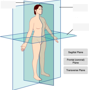
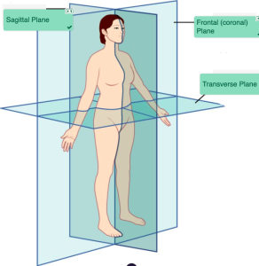
Body Planes Diagram (Text Version)
This illustration activity shows the human body standing upright in the anatomical position. Three anatomical planes are illustrated with transparent lines identifying the location of the three planes: the _______[Blank 1] which divides the left and right side of the body, the _______[Blank 2] dividing front and back portions of the body, and the _______[Blank 3] dividing top and bottom portion of the body.
Check your answers: [1]
Activity source: Medical language with the Context of Anatomy and Physiology Body by Tiffany Hunt, illustration from Anatomy and Physiology (OpenStax) licensed under CC BY 4.0./ Text version added.
Body Cavities
Use the anatomic reference system to identify the body cavities:
Body Cavities (Text Version)
- Abdominopelvic
- Abdominal
- Thoracic
- Pelvic
- Cranial
- Ventral
- Vertebral
- Diaphragm
- Dorsal
- Cranial
- Vertebral
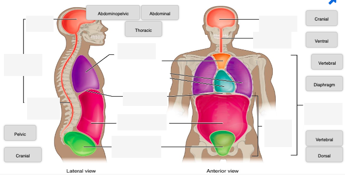
Body Cavities Diagram (Text Version)
This diagram activity shows two views of the human head and torso. The right image shows the lateral view, and the left image shows an anterior view of the human head and torso. The image is highlighting the several cavities or hollowed out spaces where organs and structures are positioned. The lateral view show four cavities: the head cavity known as the _______[Blank 1], the upper back cavity known as the _______[Blank 2], the lower back area known as the _______[Blank 3], and the chest cavity known as the ______[Blank 4] cavity. Adjoining lines for both views identify the ________[Blank 5] which is a muscular partition dividing the lungs from the location below it known as the _________[Blank 6] and followed by the lower _______[Blank 7]. The anterior view identifies the _______[Blank 8], _____[Blank 9], ________[Blank 10],________[Blank 11] cavities, and sections.
Check your answers: [2]
Activity source: Medical language with the Context of Anatomy and Physiology Dorsal by Tiffany Hunt, illustration from Anatomy and Physiology (OpenStax) licensed under CC BY 4.0./Text version added.
Directional Terms
Practice pronouncing and defining the following directional terms:
Medical Language with the Context of Anatomy and Physiology Directional Terms (Text version)
- Cranial Cavity
- KRĀ-nē-ăl kah-vi-tē
- The space formed inside the skull that the brain occupies.
- Homeostasis
- HŌ-mē-ō-STĀ-sĭs
- Biological process that results in stable equilibrium
- Posterior (or Dorsal)
- pŏs-TĒ-rē-or(Original Term)
- Describes the back or direction toward the back of the body.
- Anterior (or Ventral)
- an-TĒR-ē-ŏr (Original Term)
- Describes the front or direction toward the front of the body.
- Deep
- DĒP (Original Term)
- Describes a position farther from the surface of the body.
- Distal
- DIS-tăl (Original Term)
- Describes a position in a limb that is farther from the point of attachment or the trunk of the body.
- Inferior (or Caudal)
- in-FĒR-ē-ŏr (Original Term)
- Describes a position below or lower than another part of the body proper; near or toward the tail.
- Lateral
- LĂT-ĕr- ăl (Original Term)
- Describes the side or direction toward the side of the body.
- Medial
- MĒD-ē-ăl (Original Term)
- Describes the side or direction toward the side of the body.
- Peritoneum
- (per-it-ŏ-NĒ-ŭm)
- Serous membrane surrounding several organs in the abdominopelvic cavity. This reduces friction between the abdominal and pelvic organs and the body wall.
- Proximal
- PROK-sĭ-măl (Original Term)
- Describes a position in a limb that is nearer to the point of attachment or the trunk of the body.
- Superficial
- SOO-pĕr-FISH-ăl (Original Term)
- Describes a position in a limb that is nearer to the point of attachment or the trunk of the body.
- Superior (or Cranial)
- soo-PĒ-rē-or(Original Term)
- Describes a position above or higher than another part of the body proper.
Activity Source: Medical Language with the Context of Anatomy and Physiology Directional Terms from Medical Terminology by Grimm et al., licensed under CC BY 4.0./Re-recording of some H5P audio by Tania Deane and David McCuaig and text version added.
Regions and Quadrants of the Body
Use the anatomic reference system to identify the regions and quadrants of the body pictured below:
Regions and Quadrants of the Body (Text Version)
- Umbilical
- Epigastric
- Left Hypochondriac
- Right Lumbar
- Right Lower Quadrant
- Right Hypochondriac
- Right Iliac
- Left Lower Quadrant
- Left Lumbar
- Right Upper Quadrant
- Left Upper Quadrant
- Hypogastric
- Left Iliac
- Diaphragm
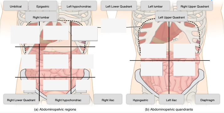
Regions and Quadrants of the Body Diagram (Text version)
This diagram activity shows two views of the abdominopelvic region. Image A shows the abdominopelvic region divided in nine sections with nine boxes. Image B shows the abdominopelvic region divided in four sections known as quadrants with a line dividing the four sections for each quadrant. For image A at the top are three white boxes identified working from right to left labeled as _______[Blank 1], followed by ______[Blank 2], and ________[Blank 3] region. The middle three boxes working from right to left identified as _______[Blank 4], followed by the _______[Blank 5], and ________[Blank 6] region. The three lower boxes working from right to left is the_______[Blank 7], followed by ________[Blank 8], and _________[Blank 9] region. Image B shows two upper white boxes and are from right to left is the _________[Blank 10] followed by the _________[Blank 11]. The two lower boxes from right to left is the _________[Blank 12] followed by _______[Blank 13].
Check your answers: [3]
Activity source: Medical language with the Context of Anatomy and Physiology Dorsal by Tiffany Hunt, illustration from Anatomy and Physiology (OpenStax) licensed under CC BY 4.0./Text version added.
Test Your Knowledge
Test your knowledge by answering the questions below:
Test Your Knowledge (Text Version)
- The smallest unit of any of these pure substances (elements) is a(n) _________ [Blank 1].
- Cell
- Atom
- Organ
- Describes a position closer to the surface of the body.
- Superficial
- Anterior
- Superior
- The plane that divides the body or organ horizontally into upper and lower portions is called the ________ [Blank 2].
- Sagittal Plane
- Transverse Plane
- Frontal Place
- The cavity that includes the cranial cavity and spinal cavity is called the _________ [Blank 3].
- Posterior cavity
- Anterior cavity
- Ventral Cavity
- The name of the layer of the membrane that covers the organs is the ________ [Blank 4].
- Serous layer
- Visceral layer
- Parietal layer
Check your answers: [4]
Activity source: Medical language with the Context of Anatomy and Physiology Reinforcement Activity by Tiffany Hunt, licensed under CC BY 4.0./Text version added.
Attribution
Except where otherwise noted, this book is adapted from Medical Terminology by Grimm et al. (2022), Nicolet College, CC BY 4.0 International. / A derivative of Building a Medical Terminology Foundation by Carter & Rutherford (2020), and Anatomy and Physiology by Betts, et al., CC BY 4.0, which can be accessed for free at OpenStax Anatomy and Physiology.
-
↵
Check your answers: Body Planes Diagram
This illustration activity shows the human body standing upright in the anatomical position. Three anatomical planes are illustrated with transparent lines identifying the location of the three planes: the sagittal plane which divides the left and right side of the body, the frontal plane dividing front and back portions of the body, and the transverse plane dividing top and bottom portion of the body. -
↵
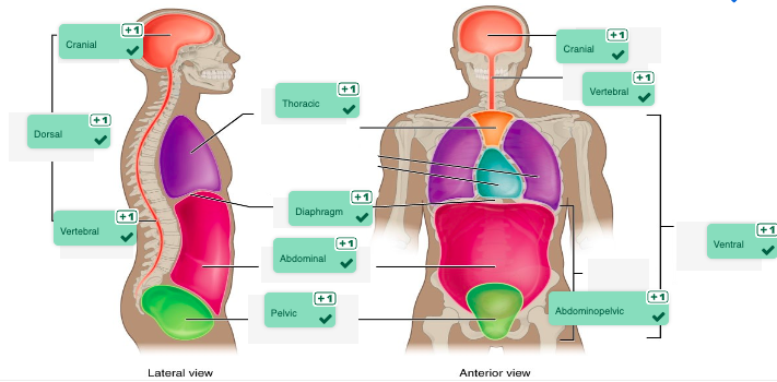
Check your answers: Body Cavities Diagram (Text Version)
This diagram activity shows two views of the human head and torso. The right image shows the lateral view, and the left image shows an anterior view of the human head and torso. The image is highlighting the several cavities or hollowed out spaces where organs and structures are positioned. The lateral view show four cavities: the head cavity known as the cranial, the upper back cavity known as the dorsal, the lower back area known as the vertebral, and the chest cavity known as the thoracic cavity. Adjoining lines for both views identify the diaphragm which is a muscular partition dividing the lungs from the location below it known as the abdominal cavity and followed by the lower pelvic cavity. The anterior view identifies the cranial, vertebral, ventral, abdominopelvic cavities, and sections. -
↵
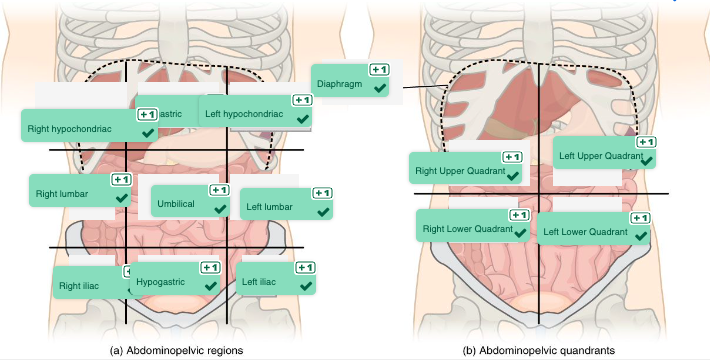
Check your answers: Regions and Quadrants of the Body Diagram (Text Version)
This diagram activity shows two views of the abdominopelvic region. Image A shows the abdominopelvic region divided in nine sections with nine boxes. Image B shows the abdominopelvic region divided in four sections known as quadrants with a line dividing the four sections for each quadrant. For image A at the top are three white boxes identified working from right to left labeled as right hypochondriac, followed by epigastric, and left hypochondriac region. The middle three boxes working from right to left identified as right lumbar, followed by the umbilical, and left lumbar region. The three lower boxes working from right to left is the right iliac, followed by hypogastric, and left iliac region. Image B show two upper white boxes and are from right to left is the right upper quadrant followed by the left upper quadrant. The two lower boxes from right to left is the right lower quadrant followed by left lower quadrant. - 1. Atom, 2. Superficial, 3. Transverse plane, 4. Posterior cavity, 5. Visceral layer ↵
