39
What is a vertebra?
Just like other delicate tissues and organs, the nervous system requires structures to protect against impact and damage. The skull encases the brain, while the vertebrae form a shield for the spinal cord. The vertebral column also provides support and flexibility to the back, allowing for a wide range of motion.
Vertebral organization:
There are 33 vertebrae, each one stacked on top of the other, which can be divided into 4 different sections. From the superior end to the inferior end of the spine, these are the cervical, thoracic, lumbar and sacral regions, each with different numbers of vertebrae. These vertebrae stacked upon one another form a continuous opening, forming what is called the vertebral column.
Each vertebra has the same general features: the vertebral foramen, which allows the spinal cord to pass through the centre of the vertebra, and the intervertebral foramina on either side of the vertebrae, which allow spinal nerves to branch out from the cord to innervate muscles throughout the body. The vertebrae themselves have function outside of just protecting the spinal cord as they also provide attachment sites for numerous ligaments and tendons – specifically the intrinsic back muscles, discussed in chapter 4. The shape, size, and density of the vertebrae vary by section based on their function.
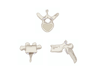
Figure 92 Vertebra bony features; superior view(top); anterior view (bottom left); lateral view(bottom right)
Vertebral organization:
The location of the vertebrae are described based on their spinal level, which is organized in a letter-number format. The letter indicates the section of the spine and the number indicates which vertebra of that section is being referred to. For instance, L5 refers to the fifth lumbar vertebra, and C2 refers to the second cervical vertebra.
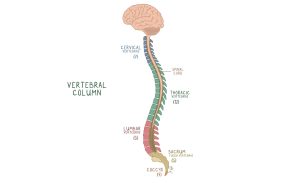
Figure 93 Segments of the vertebra organized by location
| Segment | Spinal levels | Location and function |
| Cervical | C1-C7 | Makes up the neck region of the spine. These vertebrae are smallest in size to maximize range of motion |
| Thoracic | T1-T12 | Situated through the rib cage, these vertebrae are much larger than cervical vertebrae. They help to anchor the ribs and protect the thoracic organs, such as the heart and lungs |
| Lumbar | L1-L5 | These vertebrae make up the lower back. They are very strong and dense, providing support and allowing us to lift things easily |
| Sacral | S1-S5 | These inferior vertebrae are fused in adults to form the sacrum to support the entire weight of the body. The sacrum also connects the vertebral column to the pelvis |
| Coccygeal | Co1-Co4 | These most inferior vertebrae fuse together to form the tailbone. The coccygeal vertebrae provide attachment for several pelvic ligaments |
Before delving into the next chapter let’s take some time to discuss what the nervous system is and break it up into manageable chunks. As discussed previously, the nervous system can be structurally categorized into the central nervous system (CNS), which serves as the main control centre for processing and integrating information, and the peripheral nervous system (PNS), which acts as the communication lines that relay signals between the CNS and the rest of the body. The components of the CNS are centrally located and include the spinal cord, brainstem, cerebellum, diencephalon, and cerebrum. The PNS comprises the cranial nerves, spinal nerves, and nerve plexuses. We will now explore these structures in more detail and connect them back to the remarkable functions of the nervous system.
