64 The nephron
What is the nephron?
The kidney is composed of thousands of nephrons. The nephron’s tube-like structure has varying levels of permeability throughout its length, allowing water and solutes to be filtered and/or reabsorbed. This enables urine to be excreted. The intricately structured and tightly regulated segments of the nephron enable it to clear waste and regulate fluid balance, blood pressure, and electrolyte homeostasis.
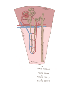
Figure 122 Zoomed image of the nephron spanning the renal medulla (lighter pink) and cortex (darker pink)
The Glomerulus:
The first structure of the nephron is the glomerulus, a cluster of capillaries branching from the afferent arteriole of the renal artery. This afferent arteriole typically maintains high blood pressure, pushing water and solutes out of the blood and into the nephron. Note that the amount of water and solutes filtered varies to achieve homeostatic concentrations, therefore filtration could be high in one moment and low in another.
The Bowman’s Capsule:
The resulting filtered fluid, referred to as the filtrate, enters the Bowman’s capsule, a double-layered epithelial structure that surrounds the glomerulus. The Bowman’s capsule acts as a selective filter which only allows certain compounds to enter the nephron to prevent blood cells or proteins from filtering in. Finding proteins or blood cells in the urine is generally a telltale sign that your kidneys are not working properly.
The Proximal convoluted Tubule:
The filtrate then flows into the proximal convoluted tubule (PCT), also located in the cortex. The PCT is lined with various microvilli to reabsorb water, sodium, glucose and other solutes back into the blood stream.
The Loop of Henle:
The filtrate then flows into the loop of Henle, which dips into the renal medulla. The segment of the nephron that extends downward, known as the descending limb, is permeable to water but not solutes, allowing water to be reabsorbed into the surrounding fluid-filled space known as the interstitium. In contrast, the ascending limb, particularly the thick segment that extends upward, is impermeable to water and actively reabsorbs sodium, potassium, and chloride to create a salt gradient in the renal medulla. This salt gradient further promotes water reabsorption in the ascending limb. The transition of the loop of Henle from the cortex and medulla allows solutes to be reabsorbed in the cortex allowing water to be reabsorbed in the medulla.
The Distal Convoluted Tubule:
The ascending limb of the loop of Henle leads into the distal convoluted tubule (DCT), which is back in the renal cortex. The DCT has fewer microvilli than the PCT and plays a role in fine-tuning sodium and calcium levels which are controlled by hormones including aldosterone and parathyroid hormone.
The juxtaglomerular apparatus (JGA) is located where the DCT comes into close contact with the afferent ateriole. The JGA acts both as a sensor of blood pressure (detecting blood pressure and solute concentration) and regulator of filtration within the kidney. In a way, the JGA functions like the kidney’s own monitoring system, able to both detect changes in blood pressure and respond to maintain homeostasis.
The Collecting Duct:
Finally, the filtrate enters the final length of the tube, the collecting duct which spans from the cortex and descends into the medulla. In response to antidiuretic hormone (ADH), these ducts adjust the final water and ion concentration of the urine, by adding water-specific channels to reabsorb water out of the kidney and back into the blood.
The Renal Papilla, Minor Calx, Major Calyx and Renal Pelvis:
The collecting ducts eventually converge at the renal papilla, where urine is funnelled into a structure called the minor calyx. The minor calyx is lined with transitional epithelium, allowing it to stretch and accommodate urine from several nephrons. Several minor calyces converge to form a larger drainage structure called the major calyx, and multiple major calyces drain into – you guessed it – an even larger structure called the renal pelvis. This gradual increase in the size of drainage structures is similar to how blood flows from capillaries to venules to veins, as thousands of nephrons eventually drain out to a single renal pelvis in each kidney.
From the renal pelvis, urine is ready to be passed into the ureters.
