24 Muscles of the Pelvic Floor
Pelvic Floor:
If you have ever reclined in a hammock, you will know there is a balance between flexibility and rigidity to ensure you are comfortable yet stable. The pelvic floor consists of several muscles and connective tissues that support the organs in your pelvis like your bladder, bowel and internal reproductive organs. These muscles help to squeeze and relax the pelvic outlet and coordinate with organs. For example, contracting and relaxing your urethra with the help of pelvic floor muscles can aid in excretion or urination. Like a hammock, the pelvic floor must be flexible to allow for adjustment, yet strong enough to maintain structure.
This subchapter will deal with the array of pelvic floor muscles and perineal muscles.
Note: Perineum refers to the area between the anus and genitalia.
Pelvic Floor Muscles:
The main group of muscles which form the pelvic floor and provide foundational support and structure are referred to as the levator ani, consisting of three distinct muscles. Because these muscles always work together, they can be considered as one.
The coccygeus muscle is a smaller, triangular muscle that lies posterior to the levator ani, and stretches from the ischial spine to the lower sacrum and coccyx. The coccygeus works in tandem with the levator ani to support the pelvic viscera.
Table 25 Muscles of the pelvic floor
| Muscle | Description: |
| Pubococcygeus | The largest muscle of the levator ani, stretching from the pubic bone to the coccyx |
| Puborectalis | A ‘U’-shaped muscle, forming a sling around the rectum. The puborectalis forms the anorectal angle – angle between your rectum and anus preventing involuntary defecation |
| Iliococcygeus | The most posterior muscle of the levator ani, running from the ischial spine of the pelvis to the coccyx |
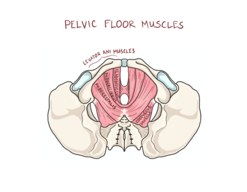
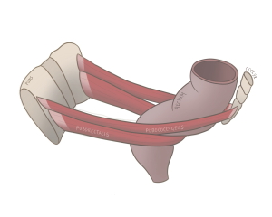 Figure 54 Lateral view of the puborectalis and pubococcygeus muscles spanning the pubis to the coccyx
The Perineal Muscles:
The perineal region is the space between the anus and the external genitalia which houses several important muscles and viscera.
Table 26 Perineal muscles
| Muscle | Description: |
| External anal sphincter | Encircles the anus and allows voluntary control over defecation |
| External urethral sphincter | Encircles the urethra which facilitates voluntary control of urination |
| Bulbospongiosus | In males, it surrounds the base of the penis and aids in erections and urination
In females, it surrounds the vaginal opening and contributes to vaginal contraction and clitoral erection |
| Ischiocavernosum | In males, it is lateral of the bulbospongiosus and helps assist the male erect penis through compressing veins
In females, it is lateral to the bulbospongiosus and assists with clitoral erection. |
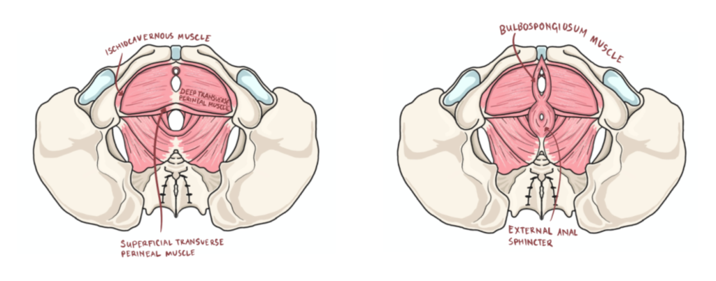
Figure 55 Inferior view of pelvic floor muscles from deep to superficial (left to right)
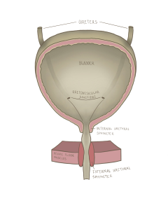
Figure 56 Coronal cross section of the bladder and urethral sphincters
Check out this meme!

Hint: This meme is referring to the important muscles covered in this chapter. While it’s easy to get lost in their long names, try breaking down these words to help you remember them!

