52 Lung Structures & Blood Supply
The Lungs
Each lung is like a tree with multiple large branches. These are the lobes that allow air to reach all parts of the organ efficiently. Each lung is divided into lobes separated by visible grooves called fissures. The left lung has two lobes (upper and lower), separated by a single oblique fissure, while the right lung has three lobes (upper, middle, and lower), divided by both horizontal and oblique fissures. These lobes allow different regions of the lung to carry out gas exchange independently. This is especially important in cases of localized damage or infection. If one lobe is compromised, others can continue functioning, much like how different branches of a tree can still gather sunlight if one is shaded.
Air continues its journey from the bronchioles into grape-like clusters called alveoli, the lungs’ true facilitators of gas exchange. Alveoli are tiny, thin-walled sacs woven tightly with capillaries. This close contact allows oxygen to pass from the air into the blood and carbon dioxide to move out of the blood to be exhaled.
Several specialized cells make this delicate exchange possible. Type I pneumocytes, which are extremely thin and flat, form most of the alveolar surface and create the direct barrier through which gas diffuses. Their thinness minimizes the distance gases have to travel, optimizing the exchange, like ultra-thin filters letting air through while keeping particles out.
Type II pneumocytes are the alveolar caretakers. Though fewer in number, they’re vital: they secrete surfactant, a slippery, soap-like fluid that reduces surface tension, preventing alveoli from sticking shut when we exhale. Without surfactant, our alveoli would collapse like deflated balloons after every breath.
In addition to these epithelial cells, macrophages patrol the alveoli like janitors on night duty, cleaning up bacteria, dust, or any particles that weren’t trapped earlier by nasal hairs or mucus in the upper respiratory tract. This keeps the alveolar lining clear and healthy.
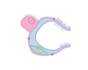
Figure 107 The alveoli and its various cell types
Blood Supply:
The lungs are constantly exchanging gases through two major pathways: pulmonary arteries and pulmonary veins. The pulmonary arteries carry deoxygenated blood from the heart to the lungs. Once oxygen has diffused into this blood in the alveoli, it becomes oxygenated and travels back to the heart through the pulmonary veins. This is one of the few cases in the body where arteries carry low-oxygen blood and veins carry high-oxygen blood, an exception to the usual rule of arteries delivering oxygen-rich blood away from the heart and veins returning oxygen-poor blood back to it.
