17 Hypothalamus and Pituitary
Starting in the brain, the hypothalamus and pituitary gland form a crucial system which produces an array of hormones. The hypothalamus acts as a manager and informs the pituitary to execute orders to remain in homeostasis. You may think of the hypothalamus as a demanding parent who is always on the case of the pituitary to finish the house chores. The hypothalamus achieves this through releasing specific hormones that either stimulate or inhibit the pituitary gland, prompting it to release its hormones to regulated key processes throughout the body.
The hypothalamus, located at the base of the brain, weighs about 4g and accounts for <1% of the brain’s total mass. The hypothalamus is directly attached to the pituitary gland via the infundibulum.
The pituitary gland (or known as the hypophysis) is a pea sized organ (0.8-1.0cm) in diameter and a weight of ~500mg. The pituitary resides within the hypophyseal fossa (commonly referred to as the Turkish Saddle): a protective indentation of the sphenoid bone of the skull. The pituitary itself is located on the midline of the head just superior to the nasal cavity and directly inferior to the optic chiasm (where the two optic tracts cross paths). Because of this location, pituitary tumors can often result in visual disturbances, particularly affecting peripheral vision.
The tandem of the hypothalamus and pituitary gland account for several significant hormones which are discussed below.

Figure 27 Sagittal cross section of the brain highlighting the optic chiasm and pituitary

Figure 28 Sagittal Cross section of the brain highlighting the pituitary and hypothalamus
This organ itself can be divided into two lobes – anterior and posterior – which are derived from different embryonic tissue. The anterior pituitary (known as the adenohypophysis) originates from the oral ectoderm and the posterior pituitary (known as the neurohypophysis) originates from the neuroectoderm directly from the diencephalon.
Hypothalamus and Anterior Pituitary:
The hypothalamus interprets concentrations of various hormones in the bloodstream and then acts on the pituitary to modulate these levels to equilibrium. The hypothalamus like an demanding manager receives information from the environment and informs the pituitary to secrete a hormone to alter the original environmental stimulus.

Figure 29 Anterior pituitary and hypothalamic hormonal products
Below is a list of hormones released by the hypothalamus, their effects on the anterior pituitary and their roles in regulating various organs throughout the body:
Table 18 Anterior Pituitary and hypothalamic hormonal pathways
| Hypothalamus secretion |
Effect on the Anterior Pituitary |
Function |
Thyrotropin-releasing hormone (TRH)
|
Thyroid stimulating hormone (TSH)
|
Stimulates thyroid to produce and release thyroid hormones which regulates metabolism, energy level and growth
|
Corticotropin releasing hormone (CRH)
|
Adrenocorticotropic hormone (ACTH)
|
Stimulates adrenal cortex to produce cortisol to regulate stress, metabolism and control blood sugar levels
|
Gonadotropin-releasing hormone (GnRH)
|
Luteinizing hormone(LH)
Follicle-stimulating Hormone (FSH)
|
LH triggers ovulation and estrogen production in females, and testosterone production in males
FSH stimulates growth and maturation of ovarian follicles in females, and production of sperm in males
|
Growth hormone-releasing hormone (GNRH)
|
Human Growth Hormone(hGH)
|
Promotes bone and muscle growth, cell repair, and metabolism
|
Prolactin-Inhibiting Hormone(dopamine)
|
Prolactin (is produced when its hypothalamic regulator is in low concentrations)
|
Stimulates milk production (lactation) in mammary glands after childbirth.
|
Hypophyseal portal system:
The hypophyseal portal system acts as the method of communication between the hypothalamus and the anterior pituitary.
The hypophyseal portal system consists of two interconnected capillary beds – one in the hypothalamus and the other in the anterior pituitary. This unique arrangement, a hallmark of portal systems allows blood carrying hormones from the hypothalamus to travel directly to the anterior pituitary without first passing through the heart. This direct pathway ensures rapid and efficient delivery of the hypothalamic hormonal messengers to their target cells in the pituitary. However, the presence of two capillary beds increases vascular resistance, leading to lower blood pressure within the system – another characteristics feature of portal systems.
The portal system can be broken up as follows:
- Hypothalamic capillary llexus: Located in the hypothalamus, this network of capillaries picks up releasing and inhibiting hormones (e.g., TRH, CRH, GnRH) secreted by hypothalamic neurons.
- Portal veins: These veins carry the hypothalamic hormones from the primary capillary plexus down the pituitary stalk to the anterior pituitary.
- Pituitary capillary plexus: In the anterior pituitary, the portal veins form another capillary bed where the hypothalamic hormones are released. These hormones act on the pituitary cells, stimulating or inhibiting the secretion of anterior pituitary hormones like TSH, ACTH, LH, and FSH.
This direct vascular link allows the hypothalamus to regulate the anterior pituitary in a highly efficient and targeted way, without the hormones needing to pass through the heart first. It’s critical for maintaining hormone balance and coordinating endocrine responses. Hormones released from the pituitary capillary plexus enter the systemic circulation, allowing them to reach their specific target organs and tissues throughout the body.
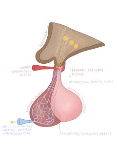
Figure 30 Hypophyseal portal system
Hypothalamus and posterior pituitary:
The posterior pituitary which receives direct nervous signals releases two hormones: vasopressin (antidiuretic hormone) and oxytocin.
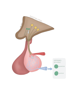
Figure 31 Anterior pituitary and hypothalamic hormonal products
- Vasopressin (antidiuretic hormone), a peptide hormone, acts on the kidneys to conserve water, and balance fluids and electrolytes.
- Oxytocin, a peptide hormone, stimulates uterine contractions during childbirth(milk-ejection reflex), and has several bonding/emotional regulating properties.
Feedback loops:
Feedback loops are regulating structures which respond to various stimuli. These feedback loops – either negative or positive – function to return variables back to homeostasis.
Negative feedback loops: A negative feedback loop is a regulatory structure which responds to a variable which is out of its homeostatic equilibrium. Negative feedback loops regulate an out-of-balance variable through inhibiting the original stimulus which triggers them. For example, the release of corticotropin releasing hormone (CRH) from the hypothalamus stimulates the anterior pituitary to release adrenotropic hormone (ACTH), which then stimulates the adrenal glands to release cortisol. When cortisol reaches a high enough concentration, it binds to the hypothalamus signalling that cortisol levels have returned to homeostasis. The hypothalamus then reduces the amount of CRH being produced which in turn reduces ACTH which depletes cortisol to homeostatic concentrations.
Figure 32 Negative feedback loop of cortisol