28 Female Pelvic Viscera
Female Pelvic Viscera
Like the male pelvic viscera, the female pelvic viscera consists of the internal organs located within the pelvic cavity, including reproductive, urinary, and digestive components.
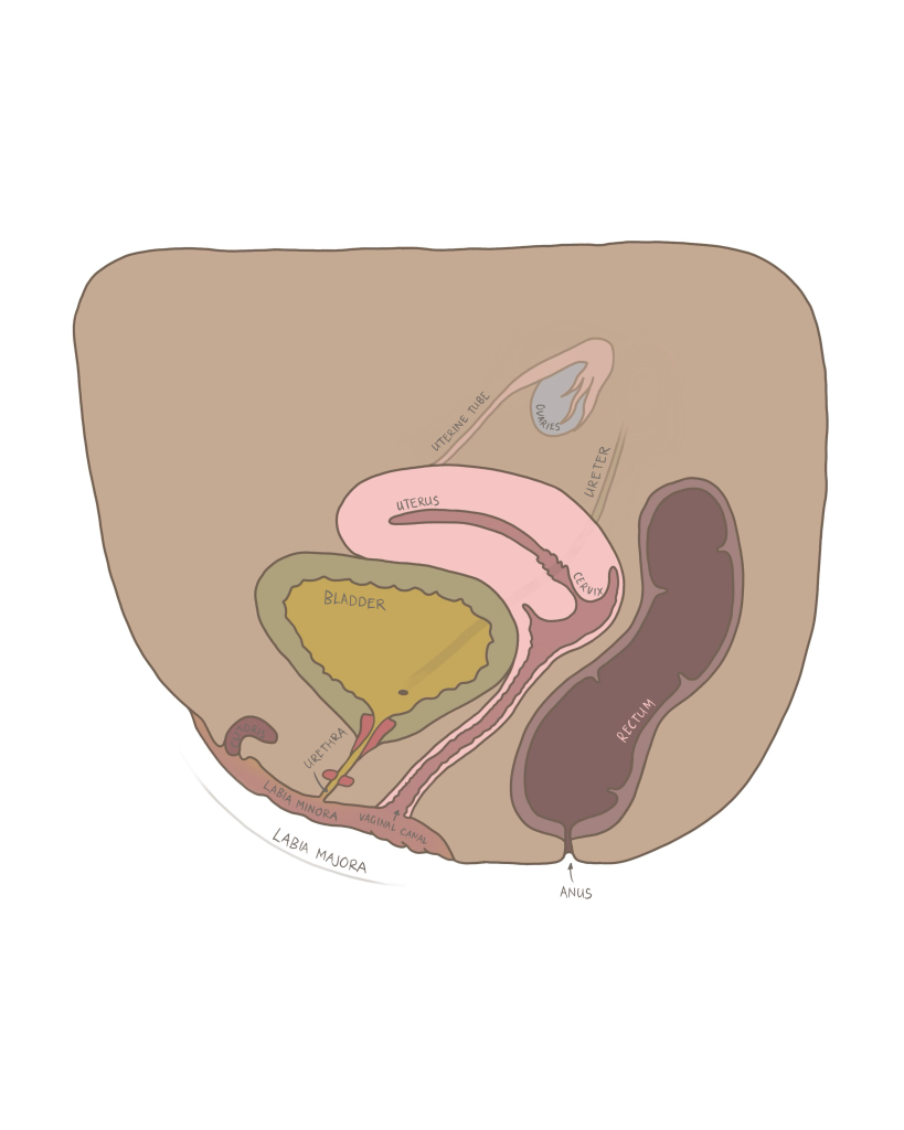
Figure 70 Sagittal view of the female pelvic viscera
Uterus:
The uterus is a hollow, muscular organ located at the midline of the pelvic cavity, situated between the urinary bladder and rectum. It has three structural regions:
Table 31 Structural components of the uterus
| Structure | Description: |
| Fundus | The dome shaped, upper portion of the uterus, located above the uterine tubes. |
| Body | The main central portion of the uterus where implantation of the fertilized egg occurs. |
| Cervix | The lower cylindrical portion of the uterus which projects into the vagina. The cervix has an internal os (opening to the uterus) and an external os (opening to the vagina). |
Uterine wall:
The uterine wall plays a key role in the implantation of a fertilized egg, and consists of three layers:
Table 32 Layers of the uterine wall
| Layer(from deep to superficial) | Description: |
| Endometrium | The inner lining of the uterus which thickens during the proliferative and excretory phases of the menstrual cycle in preparation for pregnancy. If implantation does not occur, this layer sheds during the menstrual phase of the cycle. |
| Myometrium | The thick, muscular, middle layer responsible for powerful contractions during labor and delivery. |
| Perimetrium | The outer serous layer which covers the uterus and produces lubricating fluid to prevent friction with the other pelvic organs. |

Figure 71 Anterior view of the uterus and its layers
Ovaries:
The ovaries were briefly discussed in Chapter 2 regarding their role as an endocrine gland, and yet their role is also paramount in the female reproductive system. The ovaries are situated lateral and posterior to the uterus. They are attached to the uterus via the ovarian ligament, and suspended in the pelvic cavity by the suspensory ligament of the ovary which contains the blood and nervous supply. After puberty and before menopause, the ovaries can produce and release a mature egg (ovum) during the menstrual cycle (ovulation). This typically occurs approximately every 28 days, but the frequency can vary greatly between individuals.
Uterine tubes:
The uterine tubes may be called fallopian tubes or oviducts. They are a pair of slender tubes where eggs captured from the ovaries are fertilized and then transported to the uterine cavity. The most distal portion of the uterine tubes (closest to the ovaries) is funnel-shaped and called the infundibulum. Within the infundibulum are finger-like projections called fimbriae which help capture the egg that is released after ovulation. Once the egg enters the infundibulum, it is guided to a wider part of the uterine tube called the ampulla where fertilization usually occurs. The fertilized egg then proceeds down the uterine tube through the isthmus where the tube and uterine cavity meet and exit into the uterine cavity to implant in the endometrium.
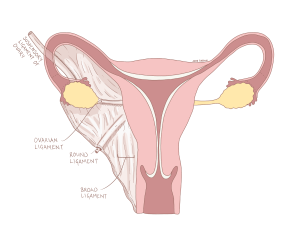
Figure 72 Anterior view of the female pelvic viscera and the surrounding ligaments
Check out this meme about the uterus!

Hint: This meme is referencing the fact that many of us are aware of the uterus as part of the menstrual cycle, but that is not the only role! The diversity of its functions is showcased in this meme.
