23 Bony Pelvis – Male and Female
The Bony Pelvis:
Like the foundation of a building, the pelvis is a load-bearing structure. Instead of having concrete and steel bearing weight, the pelvis is made of bones and ligaments. The pelvis helps protect the reproductive organs and viscera. The two hip bones that make up the pelvic girdle are joined by the sacrum, the most inferior portion of the vertebrae, completing the pelvis. Each hip bone is composed of three bones which are completely fused together in adults.
The three fused bones are:
Table 22 Bones composing the bony pelvis
| Bone | Description: |
| Ilium | The most superior portion of the pelvis. The superior part of the ilium forms the iliac crest which ends anteriorly forming the anterior superior iliac spine. |
| Ischium | The inferior and posterior part of the bony pelvis which connects to the pubis to form the obturator foramen, an opening through which vasculature and nerves flow. |
| Pubis | The anterior and inferior part of the pelvis, which fuses together with each other to form the pubic symphysis. |
In order to form the complete, ring-like structure known as the bony pelvis, the sacrum connects the two hip bones. The sacrum is a fusion of five sacral vertebrae (S1-S5), positioned at the posterior end of the pelvis. The bony pelvis supports the weight of the upper body and provides attachment points for the lower limbs. View the diagram below to get a visual representation of the hip bones:
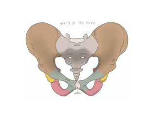
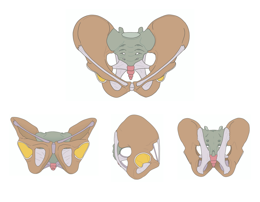
Figure 47 Anterior view (top), Inferior view (bottom left), Lateral view (bottom middle), Posterior (bottom right) of pelvis
Pelvic Foramina:
The bones and ligaments of the pelvis come together to form several openings known as foramen:
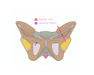
Figure 48 Anterior view bony pelvic foramen
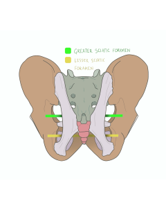
Figure 49 Posterior view of bony pelvis foramen
Table 23 Pelvic foramina and their descriptions
| Foreman | Description: |
| Inguinal canal (red line) | A passage in the lower abdominal area located above the inguinal ligament which connects iliac spine to the pubis, allowing for the passage of the spermatic cord in males and the round ligament for females (more on this later in chapter 3) |
| Obturator foramen (purple line) | The largest foramen in the body, located between the pubis and ischium covered by the obturator membrane. This allows the obturator nerve, artery and vein to pass through into the thigh which service the medial portion of the thigh |
| Greater sciatic foramen (green line) | Situated between the ilium and ischium; formed by the sacrospinous ligament, this allows the sciatic nerve – the largest nerve in the body – to pass from the pelvis to the gluteal region and down the leg |
| Lesser sciatic foramen (yellow line) | Located just below the greater sciatic foramen, allows the passage of the pudendal nerve, and internal pudendal blood vessels which supplies the genitalia and perineum |
Pelvic Brim (Pelvic Inlet):
The pelvic brim forms the pelvic inlet (opening) to the pelvic cavity and divides the true pelvis (below the brim) from the false pelvis (above the brim). The pelvic brim is approximately heart-shaped in males and oval in females.
The false pelvis supports your abdominal organs which would otherwise put immense pressure on the pelvic floor.
The true pelvis is the cavity which contains the pelvic organs. In females, the walls of the true pelvis form the bony limits of the birth canal during delivery. The opening at the bottom of the true pelvis is the pelvic outlet.
Below represents the pelvic brim and the true and false pelvises:
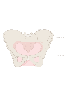
Figure 52 Anterior View of true and false pelvis
Pelvic Sexual Dimorphism:
While both male and female pelvises share the same bones and basic structure, there are several key differences, such as shape, bone density, and other features, which can generally differentiate males from females.
Table 24 Sexual Dimorphism features of the Pelvis
| Key difference | Male Feature | Female Feature |
| Bone density | Higher bone density to hold more weight | Lower bone density to account for less weight |
| Shape | Heart shaped pelvic girdle; Narrow; adapted for strength | Oval shaped pelvic girdle; Broader; adapted for childbirth |
| Subpubic angle | Less than 90º; ensuring the pelvic is narrow and compact | Greater than 90º; ensuring the pelvis is wide enough for childbirth |
| Ischial spines | Closer together to account for greater support and strength | Wider apart to accommodate delivery |
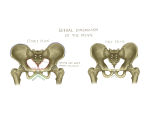
Figure 53 Anterior view of female and male pelvis
Female Pelvis:
The female pelvis is designed to carry out all the functions of the male pelvis as well as facilitate childbirth. Passage of the baby necessitates a larger, rounder pelvic inlet and outlet.
Male pelvis:
The male pelvis is solely designed for support, locomotion, and mobility. Males typically have thicker, denser bones than females and this is similarly true in the pelvis. The male pelvis can withstand more weight and bear the greater force exerted by larger muscles typically found in males.
Check out this meme!

Hint: This meme is referencing the fact that many perceive pelvic anatomy to be complicated, while the bony pelvis is composed of only three bones. Looks like those assumptions don’t have a leg to stand on!
