10 Accessory Organs: Liver, Gall Bladder, Pancreas
Accessory organs:
In a car factory there is the main assembly line which manufactures cars using parts and bolts, yet even the best factories require oil and lubricants to ensure the parts are assembled efficiently. Similarly, The accessory organs while not directly included in the alimentary canal (pathway of food and chyme), are integral to the breaking down of large food contents. The accessory organs including the liver, gallbladder and pancreas aid in digestion through numerous secretions.

Figure 15 Anterior view of accessory organs
Liver:
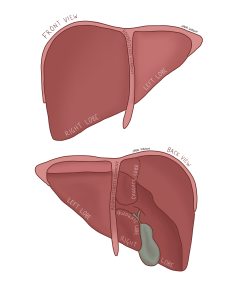
Now, we reach the body’s big, busy recycling plant. The liver, the second heaviest organ, lies in the upper right quadrant of the body. It is divided into two separate lobes connected by the falciform ligament. The liver itself can be divided into four differently sized lobes as depicted in figure 18: left, right, caudate(medial and superior) and quadrate(medial and inferior).
You will notice the liver’s inferior surface has a concave appearance, which is due to the hepatic flexure to make room for the ascending colon and houses the gallbladder:

Figure 16 Anterior view(top) and posterior view(bottom) of the liver
The liver acts like a jack-of-all-trades, serving many different functions, such as:
Table 10 Functions of the liver
| # | Description |
| 1 | Produce bile |
| 2 | Produce various proteins |
| 3 | Produce cholesterol and special proteins to help carry fats through the body |
| 4 | Convert excess glucose into glycogen |
| 5 | Regulate blood levels of amino acid |
| 6 | Process hemoglobin |
| 7 | Convert ammonia to urea to be excreted in urine |
| 8 | Clear blood of drugs |
The liver itself receives two streams of blood:
- Oxygenated blood from the hepatic artery
- Nutrient rich, deoxygenated blood from the hepatic portal system
These two streams converge in the liver in liver sinusoids to create the hepatic vein which flows into the inferior vena cava.
Gallbladder:
The gallbladder, a small greenish coloured organ inferior to the liver, has the responsibility of storing and concentrating bile produced from the liver.

Figure 17 Anterior view of the gall bladder
Imagine cleaning a greasy pan with only water; the grease will not be cleaned off, you need soap! Like dish soap, bile can emulsify fatty acids, breaking them down into smaller droplets which can be processed easier. When you are eating, your gallbladder will contract and release bile into the duodenum to break down fatty acids.
Interestingly, bile’s main pigment, bilirubin which when broken down in the small intestine releases stercobilin which gives feces its brown colour.
Pancreas:
Think of the pancreas as an unsung hero in the digestive system — while not the star onstage, they serve a vital role in the backstage crew, and without them, the show can’t go on.
The pancreas, a retroperitoneal (refers to behind the peritoneum), elongated organ does not directly receive any chyme but produces exocrine and endocrine secretions. Exocrine refers to secretions going through ducts or ampullas onto body surfaces, while endocrine refers to substances directly entering the bloodstream.
The pancreas itself can be broken down into three separate areas:
Table 11 Structural segments of the pancreas
| Segment | Description |
| Head | The widest and more rightward portion of the pancreas |
| Body | The mid-section |
| Tail | The tapered left side which leads to the spleen |

Figure 18 Anterior view of the pancreas and duodenum
The pancreas releases several exocrine products to enter the pancreatic duct into the common bile duct, and finally through the sphincter of Oddi, yet its path is a bit more complex, as depicted in fig x. These secretions are bicarbonate-rich to neutralize the highly acidic stomach contents.
With regards to the pancreas’s structure, ~99% acini cells create the exocrine secretions, and ~1% of cells known as islets of langerhans can be divided into two separate cell types:

Figure 19 Anterior view of the pancreas and microscopic depiction of pancreatic cells
Table 12 Pancreatic cell types
| Cell name | Function |
| Acinar cells | Acini cells encompass the majority of the pancreatic cells and produce most exocrine secretions which aid in digestion |
| Islets of Langerhans: | The Islets of Langerhans constitute 1-2% of the pancreas and are more relevant in homeostasis and endocrine function(chapter 2) |
Check out this meme!

Hint: The liver is like a “jack of all trades” encompassing numerous responsibilities as depicted in the accessory organ subchapter. If you need a refresher click here:

