58 The Heart
The Heart:
At a central train station, there are numerous waiting areas or platforms where passengers gather before boarding their trains and heading to their destinations. Similarly, the four chambers of the heart – right atrium, left atrium, right ventricle, and left ventricle – can be thought of as platforms where blood waits momentarily to be shuttled throughout the heart. Just as ticket gates regulate the flow of passengers between platforms and trains, the heart’s valves, namely the tricuspid, pulmonary, mitral, and aortic, control the direction of blood flow. The four chambers and four valves are integral to combining pulmonary and systemic circulation to ensure oxygenated blood arriving from the pulmonary circuit can be distributed through the systemic circuit, allowing us to exchange oxygen to our bodies.
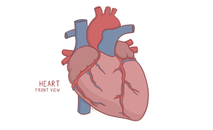
Figure 112 Anterior view of the heart
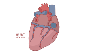
Figure 113 Posterior view of the heart
| Structures of the heart | Description of blood flow |
| Right atrium | Receives deoxygenated, venous blood returning from the systemic circuit, to be sent to the right ventricle |
| Right ventricle | Pumps deoxygenated blood through the pulmonary arteries to the lungs for gas exchange |
| Left atrium | Receives oxygen-rich blood from the four pulmonary veins to be sent to the left ventricle |
| Left ventricle | Pumps oxygen-rich blood to the rest of the body through the aorta |
| Tricuspid valve | Also known as the right atrioventricular (AV) valve, it prevents backflow from the right ventricle to the right atrium. |
| Pulmonary valve | Prevents backflow between the right ventricle and the pulmonary arteries |
| Mitral valve | Also known as the left atrioventricular (AV) valve, it prevents backflow from left ventricle to the left atrium |
| Aortic valve | Prevents backflow from the aorta to the left ventricle |
Blood returning from the systemic circuit enters the right atrium, low in oxygen and high in carbon dioxide, as oxygen has been used by tissues and converted into carbon dioxide. The CO2 has to be exchanged in the lungs for O2. To manage this, the blood moves from the right atrium into the right ventricle through the right atrioventricular, or tricuspid, valve. The right ventricle then pumps the blood through the pulmonary valve to the pulmonary trunk. The pulmonary trunk exits the right ventricle, bifurcating into the left and right pulmonary arteries. These arteries branch into smaller vessels leading to the pulmonary capillaries surrounding alveoli, where gas exchange occurs. Oxygenated, low carbon dioxide blood then flows into pulmonary veins, eventually returning to the left atrium completing the pulmonary circuit.
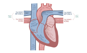
Figure 114 Anterior view of Coronal cross section of the heart
Cardiac Conduction:
In a system as intricate as the heart, a strong, motivated station master, the sinoatrial (SA) node, is essential. The SA node is a cardiac pacemaker that sets the rhythm of each stroke of the heart by generating an electrical signal that stimulates the atria to contract. The signal continues down the heart and reaches the atrioventricular (AV) node, which plays the role of an attentive engineer. Instead of rushing things immediately, the AV node double-checks that the teams are prepared before handing off the next task. This delay ensures that the ventricles can fully fill. The signal then travels down the bundle of His and its branches, acting as the team leads, who finally transmit the signal down to the Purkinje fibres, the employees who ensure synchronized contraction of the ventricles.
