54 Muscles of Respiration
What are pressure differentials?
Syringes work by using a plunger to create pressure differentials – differences between the pressure inside the machine and outside the machine. When you pull the plunger of the syringe it increases the volume in the barrel creating low pressure so air or liquid rushes in.
Inhalation
Though the human body does not have a plunger like a syringe, a series of muscles accomplishes the same effect by expanding the chest cavity and lowering the pressure inside the lungs enough to facilitate inhalation.
As you take a big inhale there are 2 key muscles which allow inhalation to occur. The diaphragm is the primary muscle of respiration. During inhalation, it contracts and flattens, increasing the volume of the thoracic cavity as the lungs fill with air. The external intercostals, which are the outermost layer of muscles between the ribs, also play a crucial role by pulling the ribs upward and outward, thereby laterally expanding the chest cavity.
For deeper inhalation (also called forced inhalation), the body recruits additional muscles to further increase the volume of the chest cavity. The sternocleidomastoid, a paired muscle located on both sides of the neck and extending from the neck to the sternum, helps lift the chest. The scalenes, a group of three paired muscles situated deeper in the neck, assist by elevating the ribs. Additionally, the pectoralis minor, a muscle in the upper chest, contributes by elevating the ribs as well.
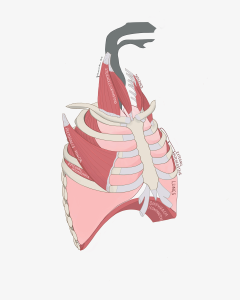
Figure 109 Anterior view of the muscles of inhalation
Exhalation
Exhalation is the process by which the body relies on several muscle groups to compress the chest cavity and expel air. At rest, the elasticity of the lungs allows air to be pushed out of the lungs.
Just as the body uses accessory muscles for deeper inhalation, it also employs additional muscles for forced exhalation. The internal intercostals, the middle layer of muscles between the ribs, pull the ribs downward and inward, reducing the volume of the chest. Within the intercostal muscles, there are three layers arranged from superficial to deep: external, internal, and innermost intercostals. The innermost intercostals are the deepest of these layers and work alongside the internal intercostals to compress the chest. The diaphragm, during exhalation, returns to its a dome shape, contributing to the compression of the thoracic cavity. The abdominal muscles play a vital role by pushing the abdominal organs upward, which forces the diaphragm to rise and helps expel air more forcefully from the lungs.
The abdominal muscles play a vital role by pushing the abdominal organs upward, which forces the diaphragm to rise and helps expel air more forcefully from the lungs.
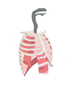
Figure 110 Anterior view of the muscles of exhalation
