51 Trachea and Bronchi
The Trachea
Imagine the trachea like a busy subway tunnel designed specifically for air traffic. As the train stops, air can both exit and enter through the windpipe like passengers. The windpipe, roughly 12 cm in length and about 2 cm in diameter, acts as the central airway that connects the upper respiratory tract to the lungs. To keep the “subway tunnel” open at all times, the trachea is reinforced with several C-shaped rings of hyaline cartilage, which act like guardrails. These rings provide both flexibility and stability, ensuring that the airway doesn’t collapse even when we breathe deeply, cough, or turn our heads.
The Bronchi
At the base of the trachea, the subway tunnel splits like a fork in the road into two large tunnels: the right and left primary bronchi. Each bronchus leads to its respective lungs, right to right, left to left. Structurally, they start off like the trachea, supported by cartilage rings. However, as these airways branch deeper into the lungs, the rigid rings slowly transition into irregular plates of cartilage and eventually give way to smooth muscle. This design is important; like adjustable tubing, the smooth muscle allows for changes in airway diameter. During an asthma attack, these muscles can constrict too much, narrowing the airways and making it difficult to breathe.
The primary bronchi continue to branch like the smaller tunnels or limbs of a tree into secondary (lobar) bronchi, each serving a specific lobe of the lung. The right lung has three lobes, so it receives three secondary bronchi, while the left has two. These, in turn, divide into tertiary (segmental) bronchi, and further into bronchioles, smaller and more numerous with each division. This intricate branching system is why the respiratory passageways are often called the “bronchial tree.”
As the “branches” get thinner, the walls lose cartilage altogether and are made entirely of smooth muscle and epithelium. The smallest of these are the terminal bronchioles, which then open into clusters of air sacs called alveoli, tiny balloon-like structures that resemble bunches of grapes. These alveoli are where the real work happens: the exchange of oxygen and carbon dioxide between the lungs and bloodstream. With over 300 million alveoli in the lungs, their combined surface area is roughly the size of a tennis court, maximizing the space available for gas exchange and keeping every cell in your body supplied with the oxygen it needs to function.
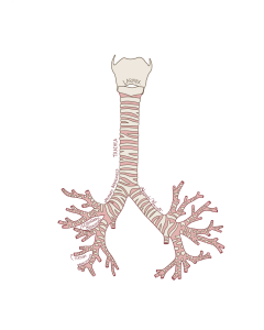
Figure 106 Anterior view of the trachea, and bronchi
