45 Meninges and Blood Brain Barrier
What are the meninges?
In addition to the skull and vertebrae, the brain and spinal cord have another shield to protect these highly complex and fragile organs called the meninges, which consists of three layers described below from superficial to deep:
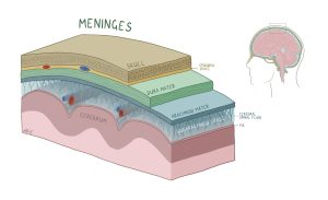
Figure 99 The layers of the meninges; dura mater (green); arachnoid mater (blue); pia mater (pink)
- Dura mater: this tough, outermost layer provides a protective barrier around the brain and spinal cord. Between the bone and the dura mater is the epidural space, containing fat and blood vessels which provide another layer of cushioning. If you’ve ever purchased something delicate online, like jewelry or a glass decorative item, you rely on the person packing your order to use multiple layers of protection to keep your item safe during shipping. The dura mater can be thought of as the cardboard box that your order ships in.
- Arachnoid mater: this middle, web-like layer lies beneath the dura mater like a spongy layer of foam insulation. The subarachnoid space lies between the arachnoid mater and a deeper layer called the pia mater. It is filled with cerebrospinal fluid for additional cushioning. Coming back to your online order, you expect that it won’t just be packed directly into a cardboard box, but rather that there is some bubble wrap to provide additional cushioning to keep your item intact and absorb the shock of any impacts. The arachnoid mater can be thought of as this bubble wrap.
- Pia mater: this innermost layer closely adheres to the spinal cord, supplying it with nutrients through blood vessels. Finally, after getting through the cardboard box and the bubble wrap, your jewelry or showpiece will likely be wrapped in a layer of felt or fabric. Like this felt or fabric, the pia mater provides the final barrier, protecting the fragile structures below.
What is the blood brain barrier?
The spinal cord and brain are some of the most important and life sustaining structures of the body and thus require the tightest security to prevent harmful blood borne pathogens from entering the nervous system, while also allowing nutrients like glucose to keep us alive. Enter the blood brain barrier (BBB), a highly selective and semi permeable barrier, separating circulating blood within the central nervous system.
The BBB is supported by three main cells:
- Endothelial cells: form the walls of the brain’s capillaries and are connected by tight junctions which restrict passage of harmful materials.
- Astrocytes: star shaped cells which support and maintain the BBB through their arm like projections
- Pericytes: embedded within the capillary walls to help regulate blood flow and provide structure
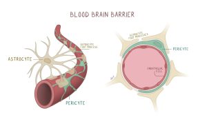
Figure 100 The blood brain barrier’s 3D representation (left); coronal cross action of blood vessels in the brain (right)
The combination of these cells aids in passive diffusion of gases like oxygen or carbon dioxide and nutrients like glucose and amino acids and help prevent passage of non-essential materials or harmful pathogens.
Cerebrospinal Fluid Production:
Mentioned before, the arachnoid mater of the meninges is filled with cerebrospinal fluid, but what is it and where does it come from? Cerebrospinal fluid (CSF) is a clear fluid which surrounds and cushions the central nervous system while providing key nutrients. CSF can be thought of as the engine oil of the brain which ensures it runs smoothly, efficiently and removes harmful waste. It is produced through networks of capillaries in brain called choroid plexuses.

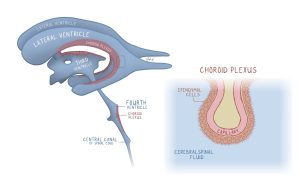
Figure 101 The ventricles of the brain (left); the choroid plexus situated within each ventricle (right)
Ventricles are cavities within the brain lined which house the choroid plexuses which produce CSF. As depicted in the diagram above, CSF production begins in the two lateral ventricles which flow into the third and fourth ventricles and into the spinal cord through the subarachnoid space of the meninges. Within the subarachnoid space there are villi-like projections, called arachnoid granulations, which can reabsorb CSF ensuring a consistent fluid pressure in the brain.
