4.4 Bones of the Appendicular Skeleton
The appendicular skeleton includes 126 bones that form the limbs, shoulders, and hips. These bones allow for movement, provide structure to the body, and protect important areas like the reproductive organs and joints.

Upper Bones of the Appendicular Skeleton
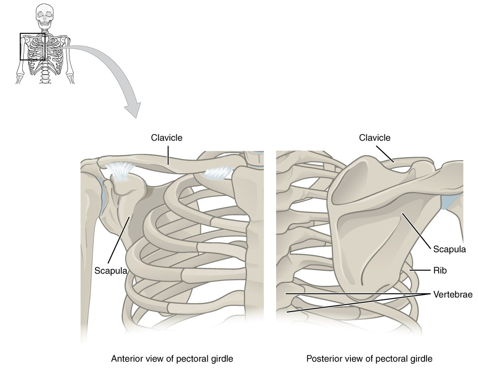
Bones of the Shoulder
- Clavicle: Known as the collarbone, it connects the sternum (chest bone) to the scapula (shoulder blade). It acts like a strut, keeping the shoulders apart and stable.
- Scapula: The shoulder blade, a large, flat, triangular bone on the upper back. It provides attachment points for many muscles and forms part of the shoulder joint.
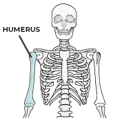
Humerus
The long bone of the upper arm connects the shoulder to the elbow. It is involved in many movements, including lifting and throwing.
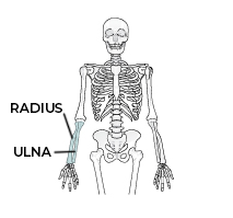
Radius and Ulna
Radius: One of the two forearm bones, located on the thumb side. It rotates around the ulna, allowing your palm to turn up (supination) or down (pronation).
Ulna: The second forearm bone, located on the pinky side. It forms the point of your elbow and works with the radius to enable wrist and arm movements.
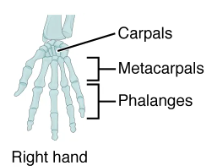
Metacarpals
The five bones in the palm of the hand connect the wrist (carpals) to the fingers (phalanges). They support hand structure and movement.
Lower Bones of the Appendicular Skeleton
The Pelvic Girdle
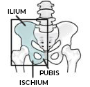
The pelvis is the large, bowl-shaped structure at the base of the spine. It connects the spine to the lower limbs and supports body weight when standing. Made of three fused bones:
- Ilium: The large, upper portion of the pelvis. You can feel the iliac crest when you place your hands on your hips.
- Ischium: The lower, back portion of the pelvis — the part you sit on.
- Pubis: The front portion of the pelvis, where the two sides join together at the pubic symphysis, a cartilage joint.
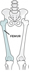
Femur
The femur, also known as the thigh bone, is the longest and strongest bone in the body. It connects the hip to the knee and supports most of your body weight while you are standing, walking, and running.
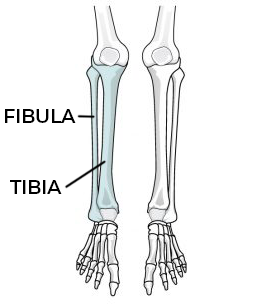
Tibia and Fibula
Tibia: Known as the shin bone, the larger and stronger of the two lower leg bones. It bears most of the body’s weight and connects the knee to the ankle.
Fibula: The thinner bone running beside the tibia. It provides support and stability but bears less weight. It also forms part of the ankle joint.
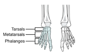
The Foot Bones
Calcaneus: The heel bone, the largest bone of the foot. It forms the base of the heel and absorbs shock during walking, running, and jumping.
Talus: Sits on top of the calcaneus and connects the foot to the tibia and fibula. It forms the ankle joint, allowing foot movement up and down.
Metatarsals: The five long bones in the middle of the foot, connecting the ankle bones (tarsals) to the toes. They help support the arch and allow for walking and balance.
