11.4 Joints: Cartilaginous and Fibrous
Cartilaginous Joints
Unlike synovial joints, which allow and support ample movement, cartilaginous joints are more restrictive. Cartilaginous joints exist where adjacent bones are joined by a firm, but flexible connective tissue known as cartilage. Cartilage provides structural support to our anatomy and helps to absorb shock or force that impacts the body.
There are two types of cartilaginous joints:
- Sychondrosis Joint: Involves bones joined together by hyaline cartilage (e.g., Epiphyseal or growth plates in long bones of growing children)
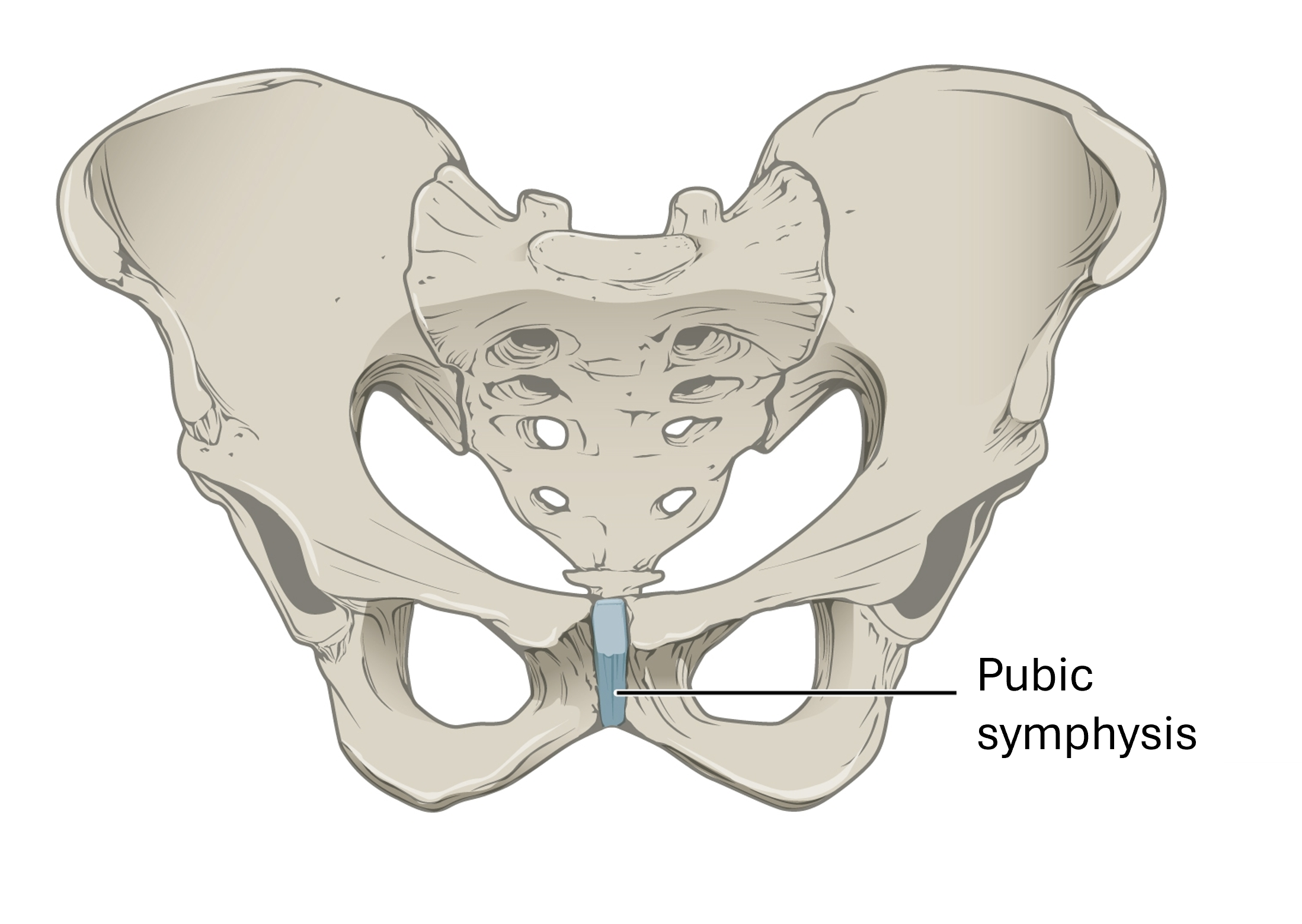
- Symphysis Joint: Involves bones joined together by fibrocartilage (e.g., Intervertebral discs of the spinal cord)
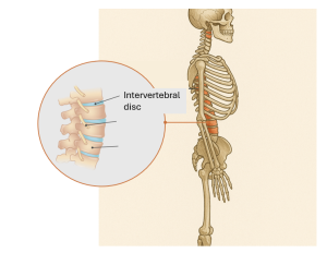
The intervertebral discs of the spinal cord. “Healthy Spine” by Injury Map, CC BY-SA 4.0 Modified: Added circle and “Skeletal side profile” image (see source below)

Fibrous Joints
Fibrous joints are made up of adjacent bones directly connected to each other by fibrous connective tissue. In fibrous joints, there is no joint cavity between bones; however, the gap between them can be narrow or wide.
There are three types of fibrous joints:
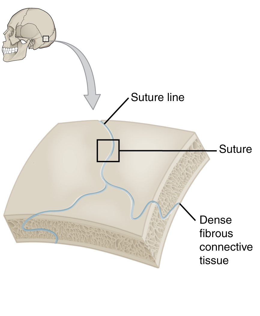
- A Suture Joint is the narrow fibrous joint found between most bones of the skull, like the cranial bone example discussed earlier.
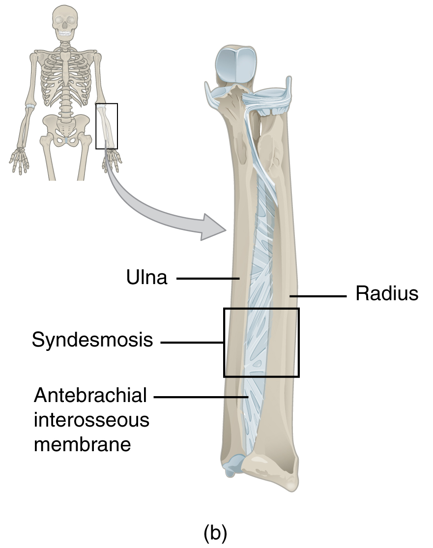
- A Syndesmosis Joint is a wider fibrous joint where bones are held together by a thin band of fibrous connective tissue (e.g., a ligament or interosseous membrane). This type of fibrous joint is found between the shaft regions of the long bones, such as the forearm or lower leg.
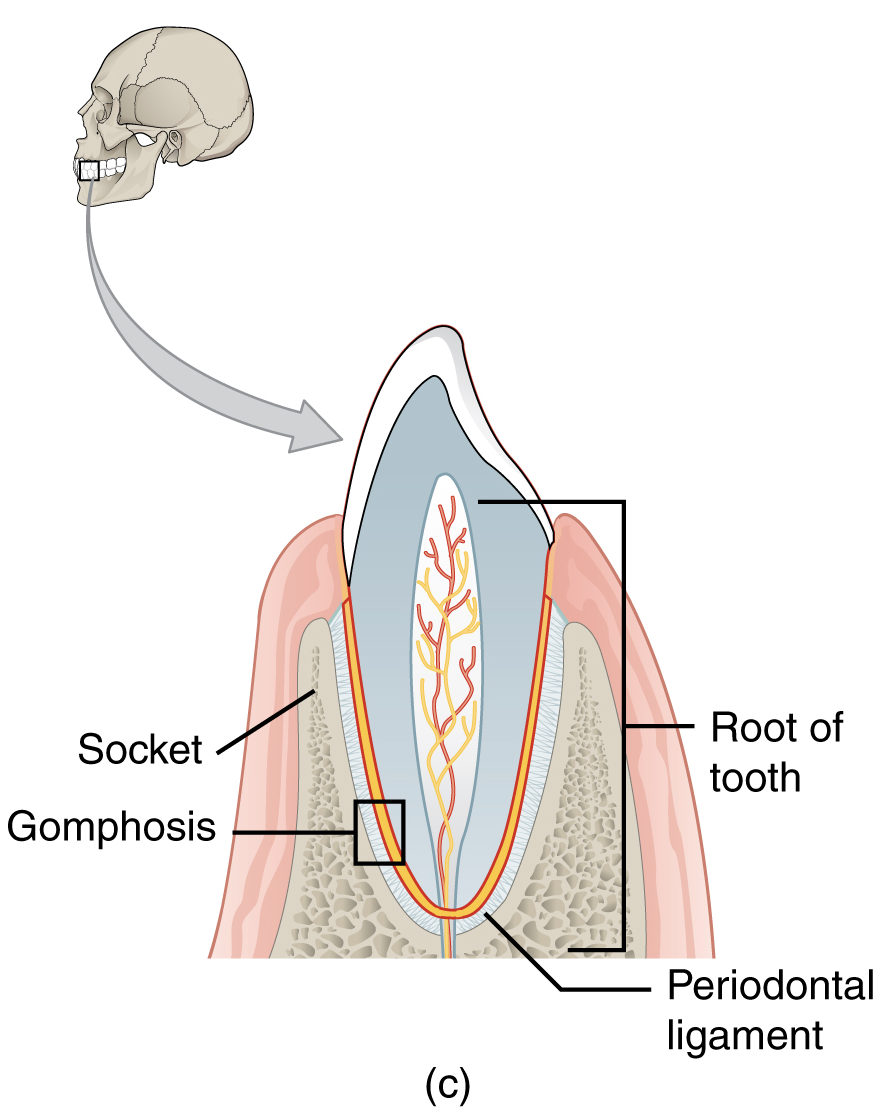
- A Gomphosis Joint is the narrow fibrous joint between the roots of a tooth and the bony socket in the jaw into which the tooth fits.
“Skeletal side profile” Image: OpenAI. (2025). ChatGPT. [Large language model]. https://chat.openai.com/chat Prompt: Create a side skeletal profile of the human body. Highlight the intervertebral discs.
