11.3 Joints: Synovial
While Chapter 4 discusses bones that make up our skeleton, it is not enough to stop there. How our skeleton functions depends not only on the bones (and muscles) that make up the body, but we also need to consider how each component is connected to the others. In addition to 206 bones, our body is held together by approximately 360 joints, connecting those bones together. Not all 360 joints, however, enable movement. Some joints are designed to permit little or even no movement. These joints provide support and even protection.
Consider your skull, for example. The part of your skull that encases the brain is known as cranial bones and provides your brain protection from external impact, and they are joined together through a specific type of joint, which does not allow movement. But what other types of joints are there?
Joint Classification
There are three classifications of joints that exist throughout the body: Synovial, Cartilaginous, and Fibrous. Each classification allows the body the amount of movement it requires: no movement, little or restricted movement, or a wide range of movement. However, the following sections will discuss all three types of joint classifications, with a greater focus on Synovial Joints because of their crucial role in movement and physical activity.
Synovial Joints
Synovial joints are the most common type of joint in the body and allow us to move (relatively) freely. Synovial joints exist where two adjacent bones are connected by a joint cavity. The joint cavity houses the ends of bones where articulation needs to occur; synovial fluid, which helps lubricate the joint; and cartilage, which covers the ends of bones to absorb shock and reduce friction. There are six types of synovial joints: the pivot, hinge, condyloid, saddle, plane (or gliding), and ball-and-socket joint.
| Joint Name | Permitted Movement | Joint Example(s) | |
|---|---|---|---|
|
1 |
Pivot |
Rotation of one bone relative to another around a single axis |
Atlantoaxial joint (Vertebrae C1-C2), Radioulnar joint |
|
2 |
Hinge |
Flexion and extension |
Elbow joint, Knee joint |
|
3 |
Saddle |
Flexion, extension, abduction, adduction and circumduction |
Thumb joint (1st Carpometacarpal Joint) |
|
4 |
Plane |
Sliding or gliding motions (limited) |
Intercarpal (Wrist) joints, Intertarsal (Foot) joints |
|
5 |
Condyloid (Ellipsoidal) Joint |
Flexion, extension, abduction, adduction and circumduction |
Wrist Joint (Radiocarpal) |
|
6 |
Ball-and-socket |
Flexion, extension, abduction, adduction, and circumduction |
Shoulder joint, Hip joint |
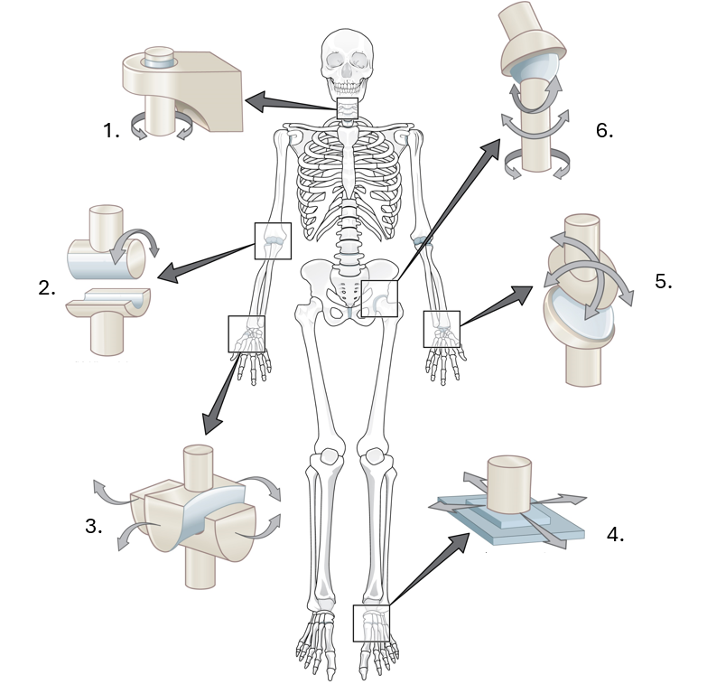
Image Description
Diagram of the human skeleton with arrows pointing to six types of synovial joints. The pivot joint at the neck allows rotational movement. Hinge joint at the elbow and knee enables bending and straightening. A condyloid joint at the wrist allows movement in two directions. The plane (gliding) joint in the ankle allows sliding movements. The saddle joint at the base of the thumb enables back-and-forth and side-to-side motion. Ball-and-socket joint at the shoulder and hip allows movement in all directions.
The variety of synovial joints in the body allows us to move in diverse ways; however, the joint capsule alone cannot support the movement our bodies demand. Tendons, Ligaments, Menisci, and Bursae all play a role in supporting joint function and movement.
-
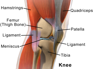
Anatomical image of the human knee showing muscles, bones, and connective tissues. Labels identify the hamstrings and quadriceps muscles, femur (thigh bone), patella (kneecap), tibia, meniscus, and two ligaments connecting the bones.”Knee Anatomy” by BruceBlaus, CC BY 3.0 Tendons: The connective tissues which connect bones to muscles and transmit force from muscle to produce movement (e.g., the patellar tendon connects the quadriceps muscle group to the tibia and plays a key role in knee extension).
- Ligaments: The connective tissue which connects bone to bone, which works to stabilize joints and limit movement (e.g., your medial cruciate ligament or MCL in your knee, restricts side-to-side movement outside of typical hinge joint flexion and extension).
- Menisci: The crescent-shaped pads of fibrocartilage found in certain joints that provide shock absorption and help distribute weight across joint surfaces (e.g. the menisci within the knee joint cushion impact during walking and running).
- Bursae: The small, fluid-filled sacs are located between bones and soft tissues that aim to reduce friction and enable smooth, painless movement (e.g., the prepatellar bursa exists in front of the patella, or kneecap, to allow smooth movement of the knee over the knee during movements such as kneeling).

Despite the support of these structures, some joints are more susceptible to injury or movement-related issues than others. Conditions that impact movement and injuries that commonly occur because of movement and sport will be discussed later in Chapter 11.9.
