80 Vision
Vision (or sight) is the ability to sense light and see. The eye is the special sensory organ that collects and focuses light and forms images. The eye, however, is not sufficient for us to see. The brain also plays a necessary role in vision. Vision is our primary sense and more than 50 per cent of the cerebral cortex is devoted to processing visual information. A person with normal colour vision can differentiate between hundreds of thousands of different colours, hues, and shades.
HOW THE EYE WORKS
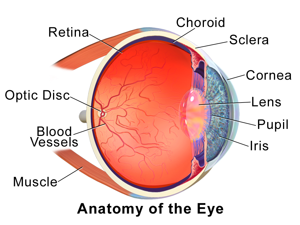
The figure below shows the anatomy of the human eye in cross-section. The eye gathers and focuses light to form an image, and then changes the image to nerve impulses that travel to the brain. The eye’s functions are summarized in the following steps.
- Light passes first through the cornea, which is a clear outer layer that protects the eye and helps to focus the light by refracting (or bending) it.
- Next, light enters the interior of the eye through an opening called the pupil. The size of this opening is controlled by the coloured part of the eye (called the iris), which adjusts the size based on the brightness of the light. The iris causes the pupil to narrow in bright light and widen in dim light. Filling the space between the cornea and the iris is a semi-gelatinous fluid called aqueous humor and functions to maintain the shape of the eye.
- The light then passes through the lens, which refracts the light even more and focuses it on the retina at the back of the eye, as an inverted image. Sitting behind the lens is a gelatinous fluid called vitreous humor, which functions to maintain the shape of the eye.
- The retina contains two types of photoreceptors: rod and cone cells . Rod cells, which are found mainly in all areas of the retina other than the very center, are particularly sensitive to low levels of light. Cone cells, which are found mainly in the center of the retina, are sensitive to light of different colours, and allow colour vision. The rods and cones convert the light that strikes them to nerve impulses.
- The nerve impulses from the rods and cones travel to the optic nerve via the optic disc (also known as the optic nerve), which is a circular area at the back of the eye where the optic nerve connects to the retina.
COLOUR VISION
Humans have colour vision because we have three types of cone cells: blue, green and red. Each of these types of cone cell detects a specific wavelength of light, for which they are named. The combined stimulus is then perceived as a specific colour, based on the ratio of the amount stimulus coming from each of the three types of cone cells. Do you know what else uses these same three pieces of information to communicate colour? Your computer monitor! When working in a creative program, such as Paint, these three reference points of red (R), green (G), and blue (B), can be used to create any of the million colours the human eye can perceive, as illustrated in Figure 8.7.5. Take a look at each of the numerical values for red, green, and blue and what colour their combined values create:
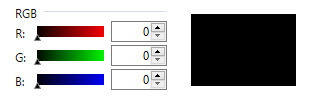
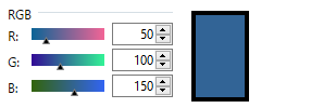
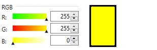
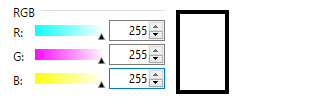
Figure 8.7.5 RGB colours.
ROLE OF THE BRAIN IN VISION
The optic nerves from both eyes meet and cross just below the bottom of the hypothalamus in the brain. The information from both eyes is sent to the visual cortex in the occipital lobe of the cerebrum, which is part of the cerebra cortex. The visual cortex is the largest system in the human brain, and is responsible for processing visual images. It interprets messages from both eyes and “tells” us what we are seeing.
VISION PROBLEMS

Vision problems are very common. Two of the most common are myopia and hyperopia, and they often start in childhood or adolescence. Another common problem, called presbyopia, occurs in most people, beginning in middle adulthood. In all three conditions, the eyes fail to focus images correctly on the retina, resulting in blurred vision.
Myopia
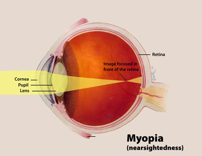
Myopia (or nearsightedness) occurs when the light that comes into the eye does not directly focus on the retina, but in front of it, as shown in Figure 8.7.7. As a result, distant objects may appear out of focus, but the focus of close objects is not affected. Myopia may occur because the eyeball is elongated from front to back, or because the cornea is too curved. Myopia can be corrected with the use of corrective lenses, either eyeglasses or contact lenses. Myopia can also be corrected by refractive surgery performed with a laser.
Hyperopia
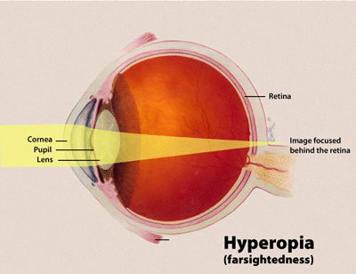
Hyperopia (or farsightedness) happens when the light coming into the eye does not directly focus on the retina but behind it, as shown in Figure 8.7.8. This causes close objects to appear out of focus, but does not affect the focus of distant objects. Hyperopia may occur because the eyeball is too short from front to back, or because the lens is not curved enough. Hyperopia can be corrected through the use of corrective lenses or laser surgery.
Presbyopia
Presbyopia is a vision problem associated with aging, in which the eye gradually loses its ability to focus on close objects. The precise origin of presbyopia is not known for certain, but evidence suggests that the lens may become less elastic with age, causing the muscles that control the lens to lose power as people grow older. The first signs of presbyopia — eyestrain, difficulty seeing in dim light, problems focusing on small objects and fine print — are usually first noticed between the ages of 40 and 50. Most older people with this problem use corrective lenses to focus on close objects, because surgical procedures to correct presbyopia have not been as successful as those for myopia and hyperopia.
Human Biology Copyright © 2020 by Christine Miller is licensed under a Creative Commons Attribution-NonCommercial 4.0 International License, except where otherwise noted.
