11 1.2 Tumour Formation Assay
Brief Overview
Experiment Protocol
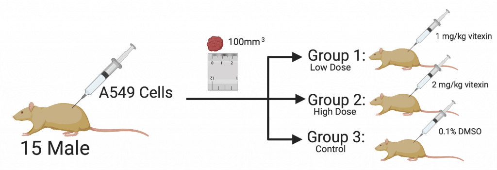
For the last part of the experiment, fifteen male nude mice aged 5–6 weeks were purchased from Shanghai Laboratory Animals Center. They injected the mice with 2×10⁶ A549 cells into a single side of the posterior flank of the mice. Every three days, they measured the size of the tumour using a calliper. When the tumour size reached approximately 100 mm3, the mice were randomized into three groups. The mice in low dose were treated with 1 mg/kg vitexin, and the high dose group was treated with 2 mg/kg vitexin while the mice in the control group received 0.1% DMSO daily for four weeks. At the end of the study (Day 19), the tumours were excised and weighed.
Results
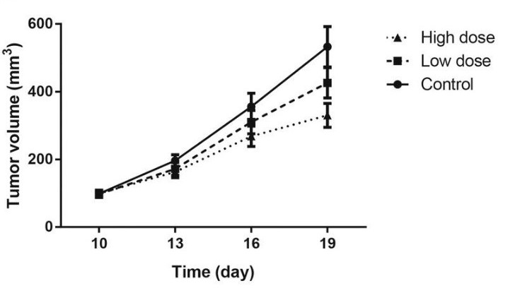
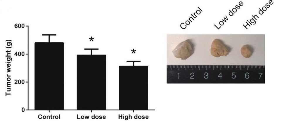
The treatment of vitexin was shown to reduce the rate of growth of the A549 cell-derived tumours. In figure 2, vitexin at higher doses was able to show an evident decrease in the rate of growth when compared to tumours not treated. When measuring the weight of the tumours after the experiment ended, there are similar results. The end weight of the tumours is significantly lower with treatment of vitexin, as seen in figure 3.
Statistical Analysis
All the statistical analyses for this experiment were performed using GraphPad Prism 6.0. All experimental data are shown as the mean standard deviation and analyzed using a one-way analysis of variance, which is a technique that can be used to compare means of two or more samples. Also, Dunnett’s post hoc test was used, and a P value less than 0.05 was considered to indicate a statistically significant difference.
Summary
The researchers conducted the following in vivo experiments to determine the effects of vitexin on tumours:
- A549 cells were injected in mice and were able to grow to form tumours.
- Tumours were treated with two different doses of vitexin.
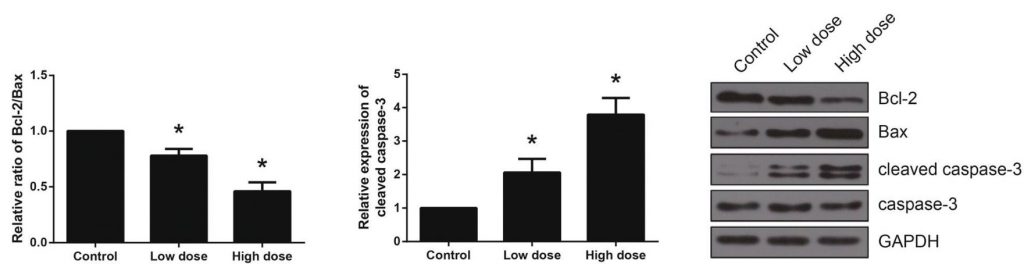
- Western blot analysis showed a decrease in Bcl-2/Bax ratio and an increase in cleaved caspase-3.
Describes a study in which the organism is outside of the living body and in an artificial environment. Adapted from Merriam-Webster.
Describes a study in which the organism is in a living body. Adapted from Merriam-Webster.