9 1.2 Vitexin and the Mitochondria
Brief Overview
Mitochondria is the powerhouse of the cell and is where ATP production occurs. Fluorescent dyes such as JC-1 staining were used to monitor the mitochondrial membrane potential (MMP). In a healthy functioning cell, the direction of the mitochondrial potential is to produce an inward current of cations and outward transport of anions to promote the buildup of cations in the mitochondria. This electrochemical gradient drives the synthesis of ATP. In a cell that is going through apoptosis, the MMP decreases as the pores of the membrane open up, and the electrochemical gradient is lost. Being able to measure the MMP is an excellent indication of a healthy versus unhealthy cell since mitochondria are inherently involved in the apoptotic process of cells.
Experiment Protocol
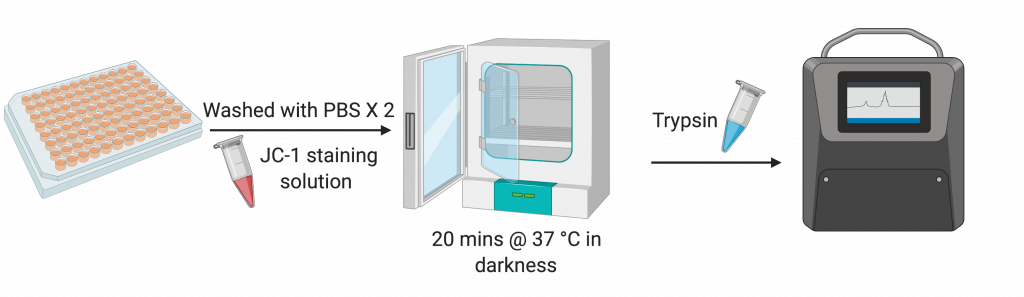
Following the initial treatment with the different doses of vitexin for 48 hours, a classical JC-1 staining method was used to determine the mitochondrial membrane potential. After the vitexin treatment, the cells were harvested, washed with PBS twice and incubated with 500 μM of JC-1 staining solution in the dark for 20 minutes in 37 degrees Celsius. Next, the authors suspended the cells with trypsin and with the aid of a flow cytometer, the cells were analyzed.
Experiment Overview
Many proteins and pathways are involved in the regulation of apoptosis, including the mitochondria. In the following experiment, Liu et al. (2019) measured the mitochondrial membrane potential (MMP) of cells to determine if vitexin can affect the functioning of mitochondria in A549 cells.
MMP is crucial to the functioning of the mitochondria. It’s the driving force for the transport of proteins and ions that keep the mitochondria functional. Even more importantly, alongside the protein gradient, it ensures that the cell can produce ATP. For these reasons, MMP changes are a good indication of a dysfunctional mitochondria.[1]
JC-1 (5,5′,6,6′-tetrachloro-1,1′,3,3′-tetraethylbenzimi- dazolylcarbocyanine iodide) is a dye that is often used to detect MMP changes. Normally, the dye is a fluorescent green, however it tends to form aggregates in mitochondria that have a high MMP, creating a red fluorescent colour. The fluorescence of cells can be detected by numerous techniques, such as spectrofluorimetry and microscopy. In this experiment, the researchers used flow cytometry.[2]
Results
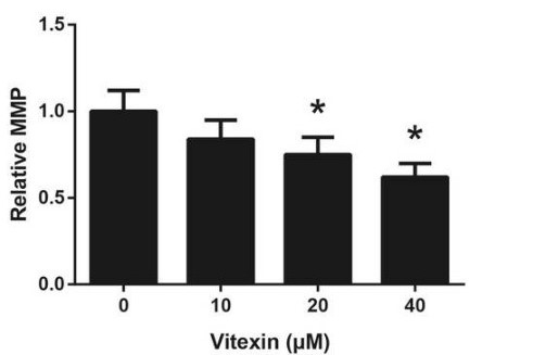
By using JC-1 staining, the researchers were able to determine what effect vitexin has on the A549 cells’ MMP. After treatment with vitexin, there was a clear loss of MMP compared to A549 cells that were left untreated. The concentration of the vitexin administered mattered too, as the effects were seen as dose-dependent.
Experiment Overview
In addition to changes to MMP, levels of cytochrome c in the mitochondria and cytoplasm can also indicate mitochondrial dysfunction. Cytochrome c is normally found close to the inner mitochondrial membrane, where it transfers electrons in the electron transport chain, leading to the production of ATP. Despite the fact that it creates the energy needed for life, it is also required for apoptosis to occur. The release of cytochrome c from the mitochondria is thought to be a result of mitochondrial dysfunction. Stress signals lead to hyperpolarization of the mitochondria, increasing MMP and producing toxic reactive oxygen species (ROS). The ROS produced have the downstream effect of causing cytochrome c release into the cytoplasm. Once in the cytoplasm, cytochrome c can induce apoptosis.[3] Liu et al. (2019) used western blot analysis in order to determine the levels of cytochrome c in the mitochondria and in the cytoplasm.
Results
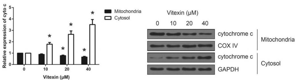
To add further support that vitexin causes mitochondrial dysfunction, the researchers analyzed cytochrome c levels using western blot analysis. Their results, as seen in figure 2, show a clear increase in cytosolic cytochrome c and a decrease in mitochondrial cytochrome c, which would indicate that cytochrome c found in the mitochondria was leaking into the cytosol. COX IV was used a control in the analysis of mitochondrial cytochrome c and GAPDH was used as a control in the analysis of cytosolic cytochrome c.
Summary
Researchers conducted the following experiment to determine what effect vitexin has on the functioning of the mitochondria of A549 cells:
- JC-1 staining to determine the change in mitochondrial membrane potential (MMP) in A549 cells.
- Western blot analysis to determine the change in cytochrome c levels in the cytosol and mitochondria.
Researchers found that vitexin treatment led to the a decrease in MMP and an increase in cytosolic cytochrome c in A549 cells.
- Mitochondrial membrane potential. [accessed 2019 Nov 24]. https://www.ncbi.nlm.nih.gov/pmc/articles/PMC5792320/ ↵
- Perelman A, Wachtel C, Cohen M, Haupt S, Shapiro H, Tzur A. 2012. JC-1: alternative excitation wavelengths facilitate mitochondrial membrane potential cytometry. Cell Death & Disease. 3(11):e430–e430. doi:10.1038/cddis.2012.171. ↵
- Hüttemann M, Pecina P, Rainbolt M, Sanderson TH, Kagan VE, Samavati L, Doan JW, Lee I. 2011. The multiple functions of cytochrome c and their regulation in life and death decisions of the mammalian cell: from respiration to apoptosis. Mitochondrion. 11(3):369–381. doi:10.1016/j.mito.2011.01.010. ↵