4 Atlas of some normal organs
These pages show various images of normal organs. This is not required reading, but it may be a helpful reference if you have only seen fixed specimens. When looking at these photos, think of how the gross appearance corresponds with what you know of the histologic structure. Also, please appreciate the range of appearances of normal organs.
Above: Normal heart, cat, cut surface after transverse slice to remove the apex. The wall thicknesses have a 3:1 ratio of left: right ventricular free walls (ignoring the papillary muscle), and 3:1 ratio of septum: right ventricular free wall. Note the colour of the myocardium and the size of the right and left ventricles (i.e. the chambers). The patchy dark red discolouration of the lung is an artefact due to postmortem pooling of blood.
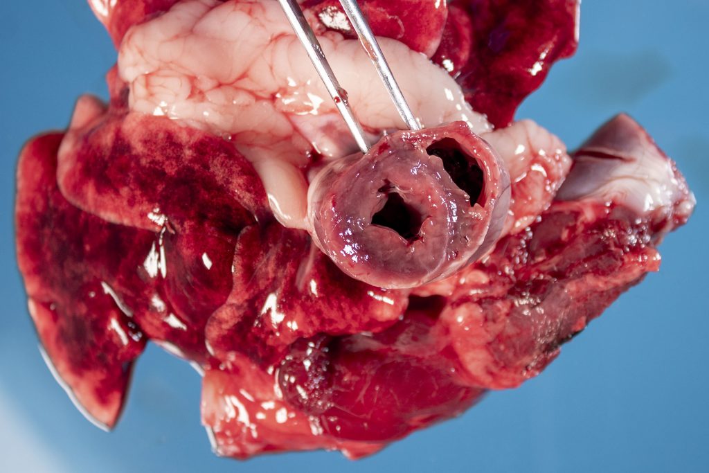
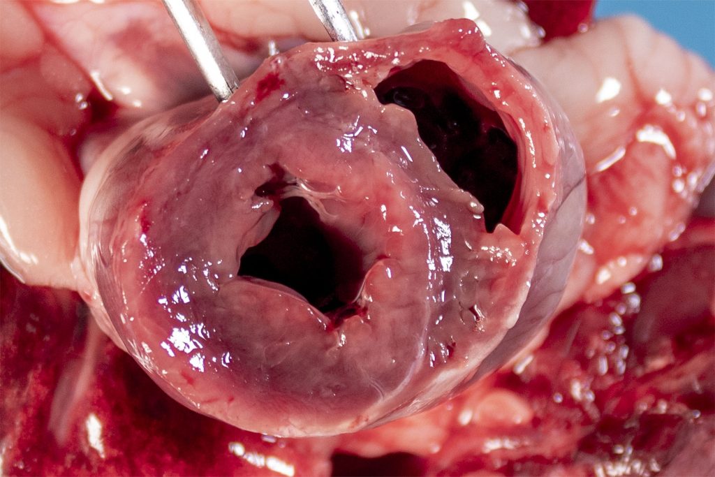
Above: Normal left atrioventricular valve, dog. Note also the left atrium (bottom) and ventricle (top).
Above: Sagittal sections of the skull from a calf.
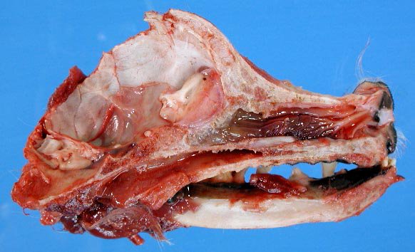
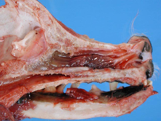
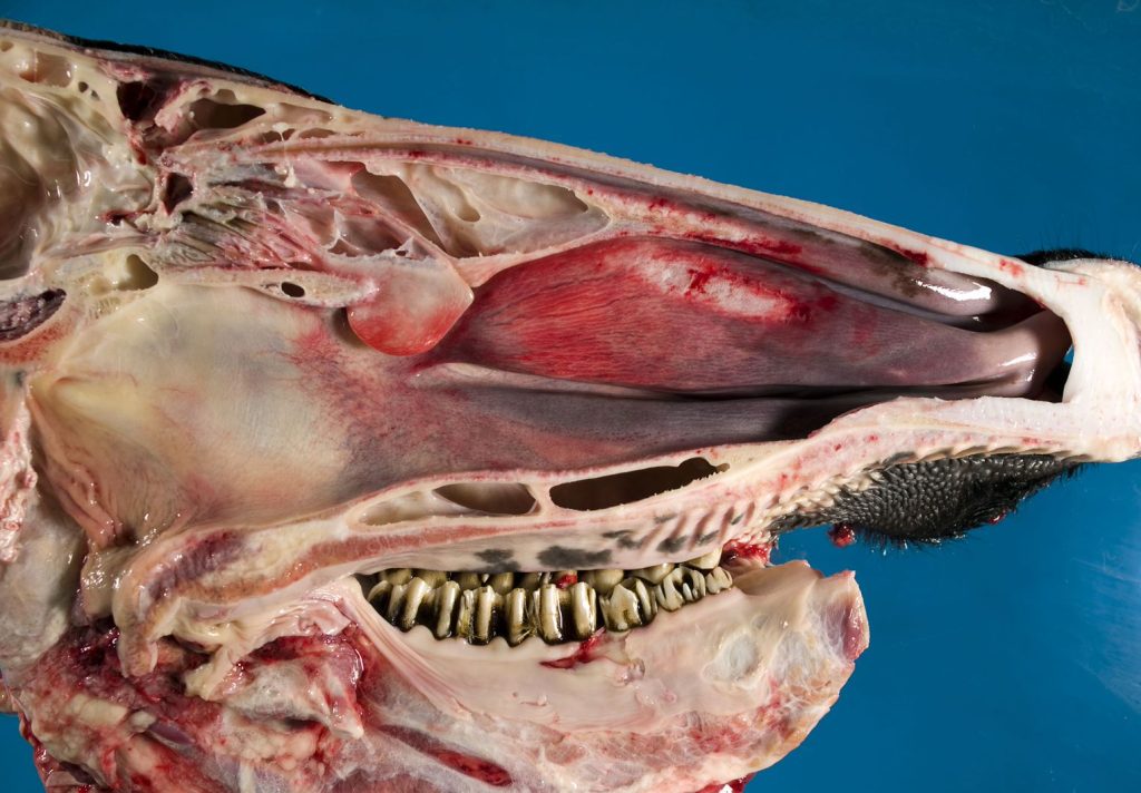
Above: Sagittal sections of the skull from a dog.
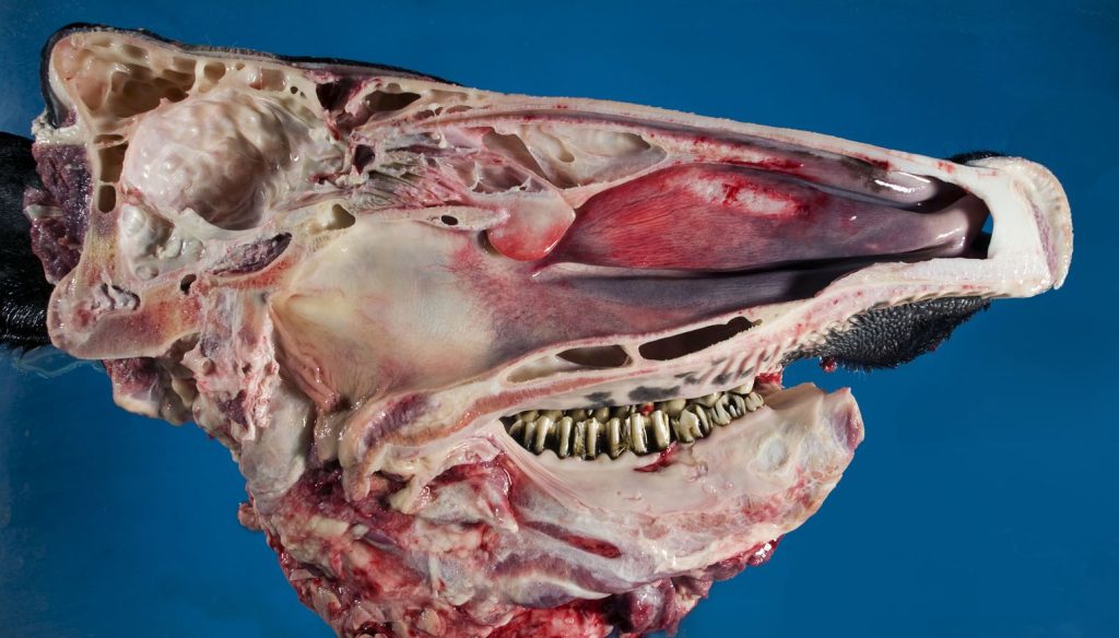
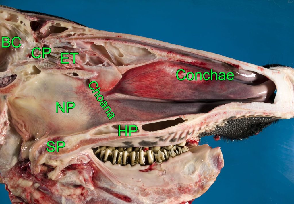
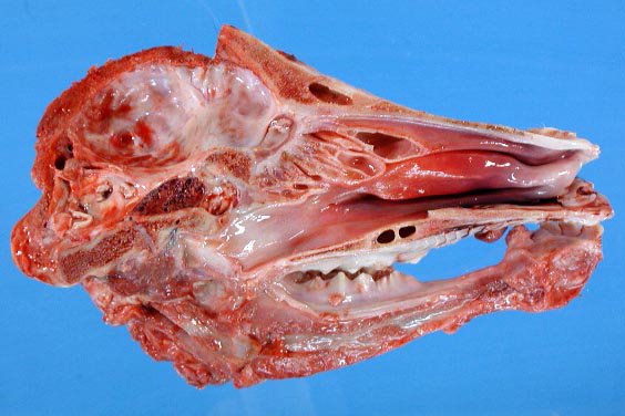
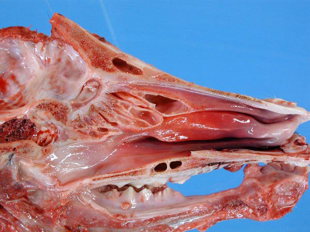
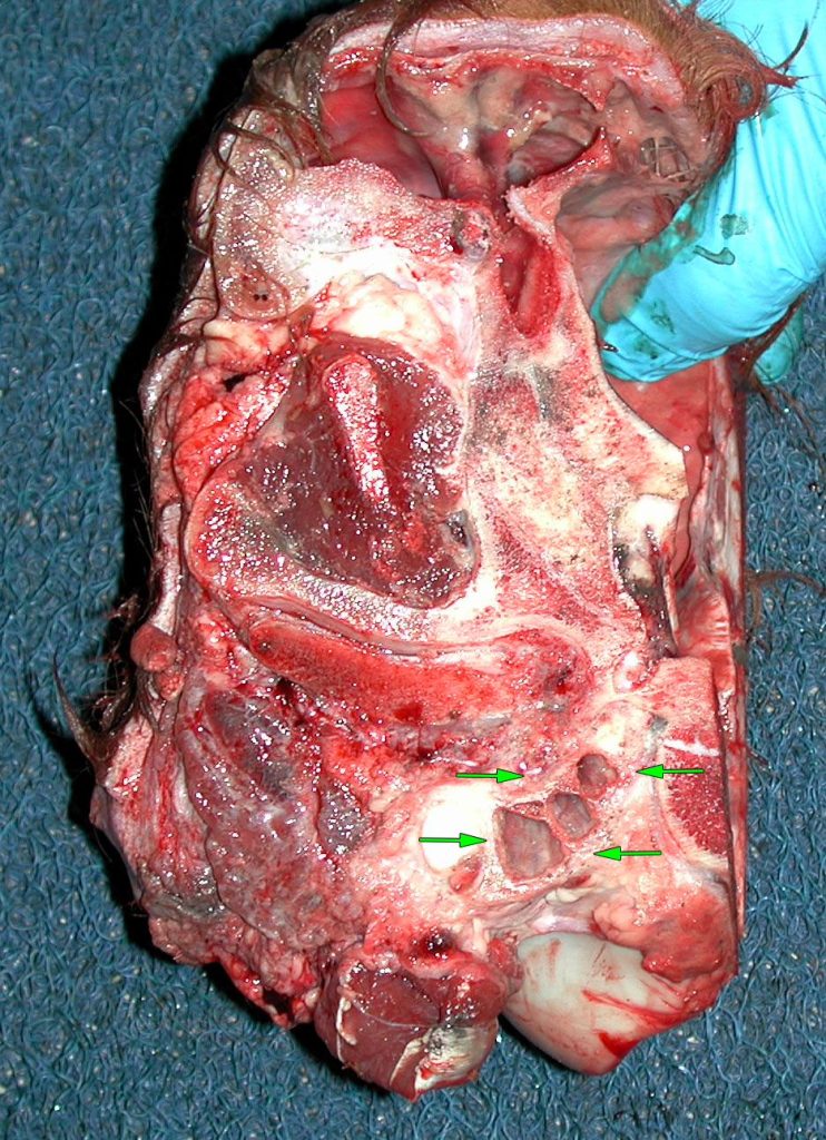
Above: Sagittal sections of the skull from a cow. The bottom photo of the cow skull is labelled with: BC, braincase; CP, cribriform plate; ET, ethmoid turbinates/conchae; maxillary conchae (turbinates); choanae; NP, nasopharynx; HP, hard palate; SP, soft palate.
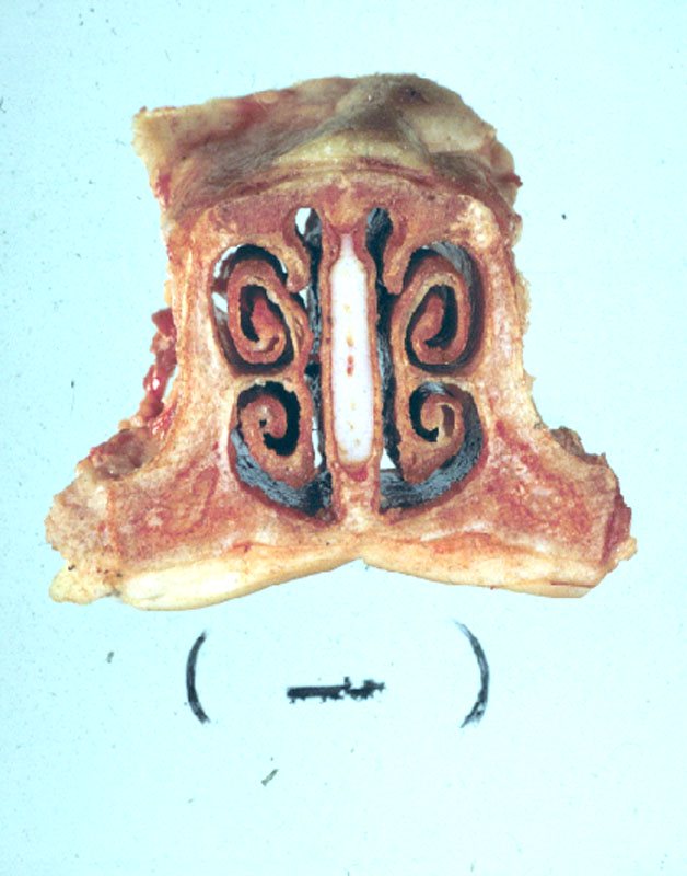
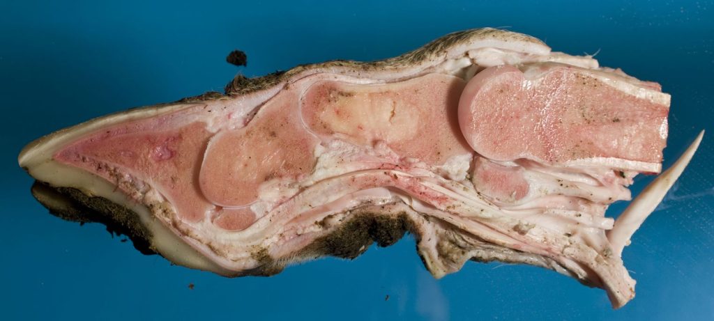
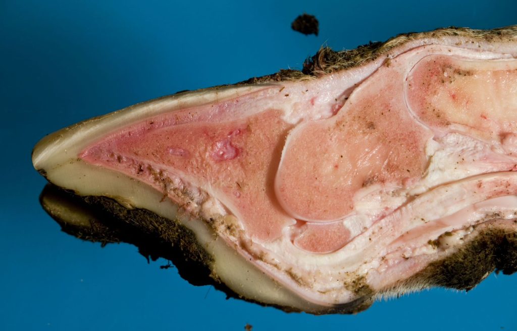
Above: Snout, pig, transverse section. Note the conchae (turbinates) and nasal septum.
Above: Tympanic bulla, steer. The head is sectioned sagitally and the brain is removed (the finger is in the braincase). The arrows show the left tympanic bulla (middle ear) within the petrous temporal bone.
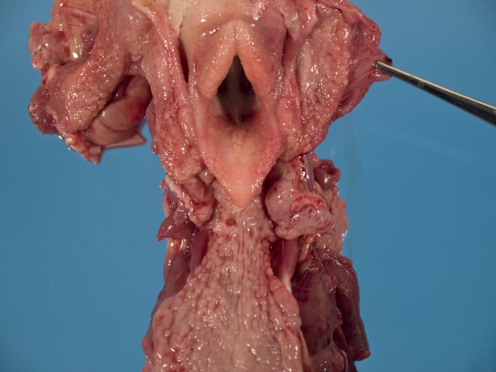
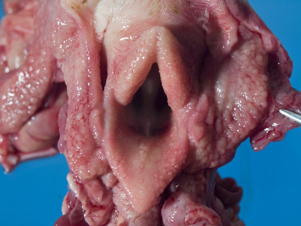
Above: Pharynx and larynx, foal. The tiny nodules in the pharyngeal mucosa represent prominent lymphoid tissue, which is normal in a young animal. The larynx is also visible, including epiglottis.
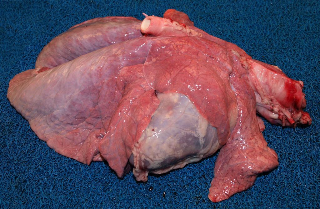
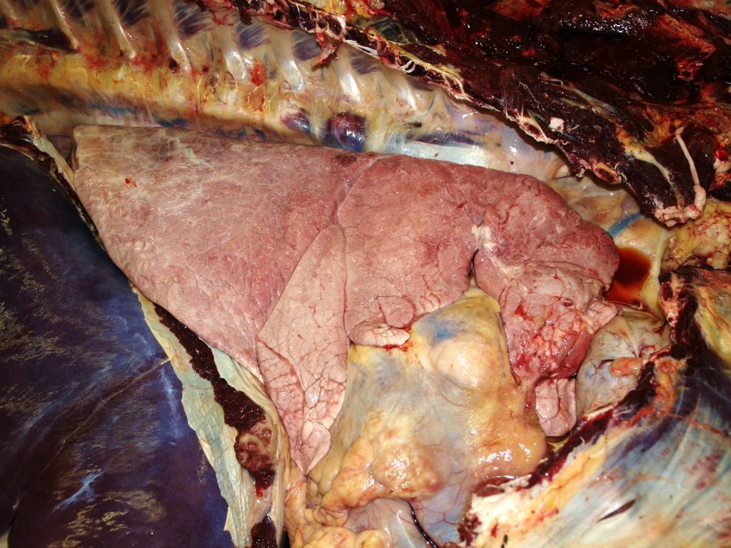
Above: Normal lung from two calves. Note also the heart, still within the pericardial sac.
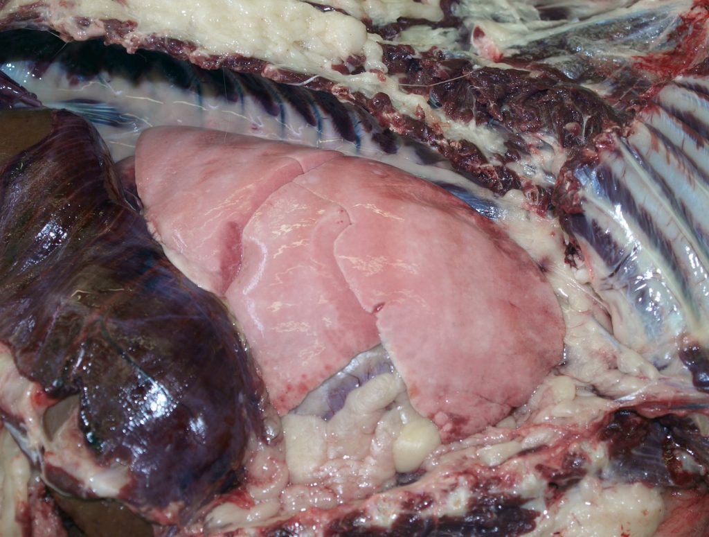
Above: Right lung from two dogs. The ribs have been removed. Note also the diaphragm, and liver.

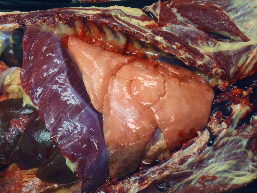
Above: Normal lung, pony. The right dorsal colon is also visible, caudal to diaphragm.
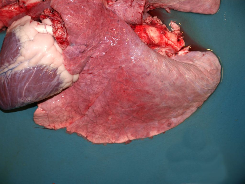
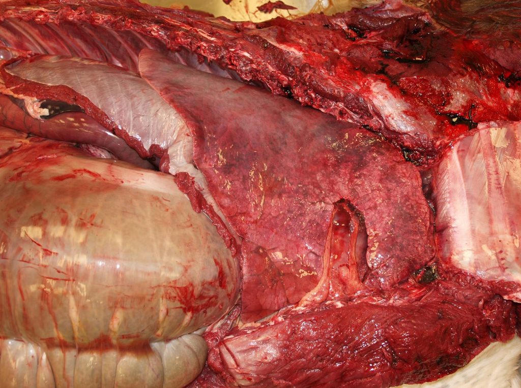
Above: Normal lung, calf. Note also the heart, with the pericardium opened, and epicardial
fat visible in the coronary and paraconal grooves.
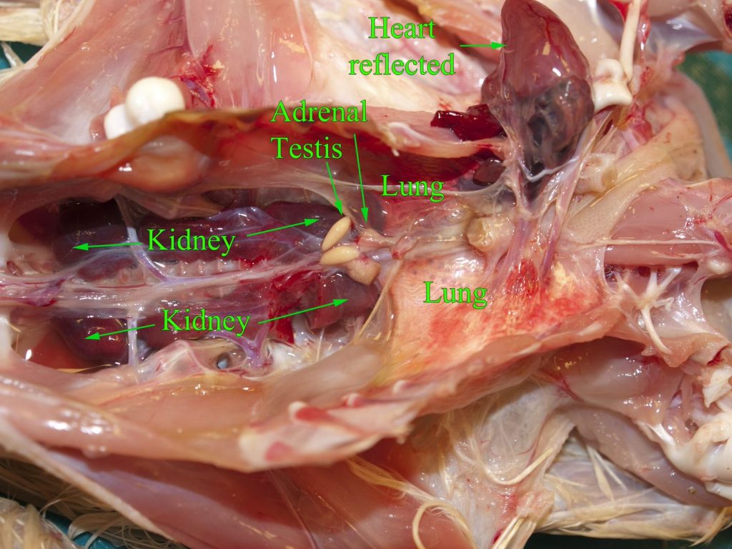
Above: Normal abdominal and thoracic viscera, chicken. The stomachs, intestines and liver have been removed.
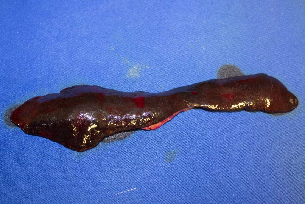
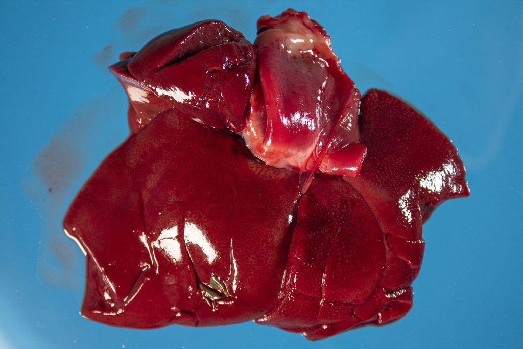
Above: Normal spleen, cat.
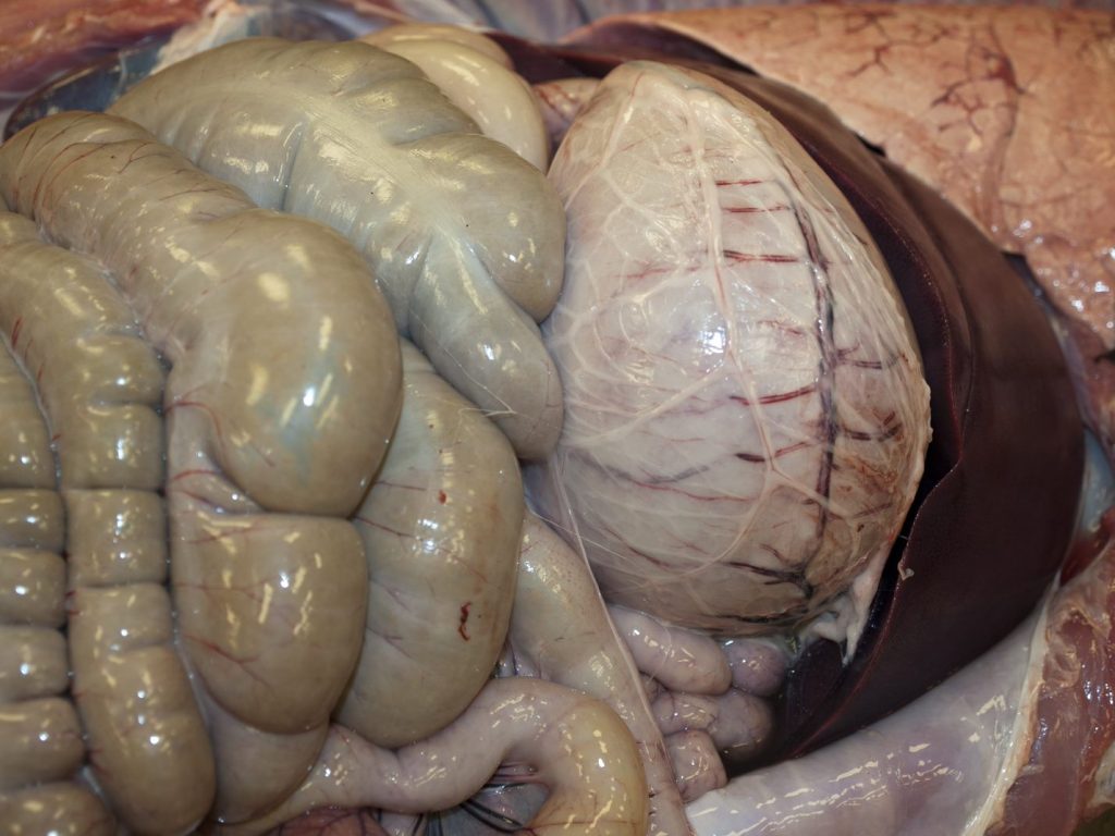

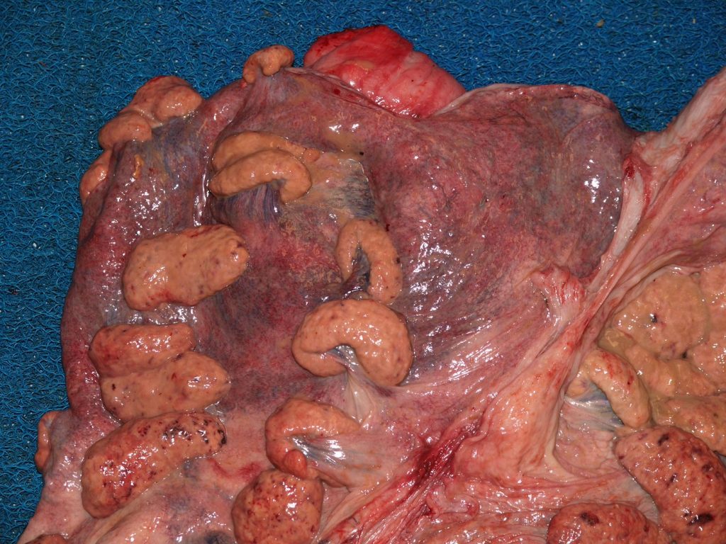
Above: Normal cranial abdomen, pig. Pigs normally have thin thread-like strands of fibrin on the serosal surface of the intestine. Visible are the liver (right), stomach (middle) and colon (left).
Above: A portion of normal stomach, mucosal surface, cat. The rugae are visible in the fundus. The pylorus is to the right and lacks rugae. A tiny piece of distal esophagus is at the far left, recognizable from its white mucosa.
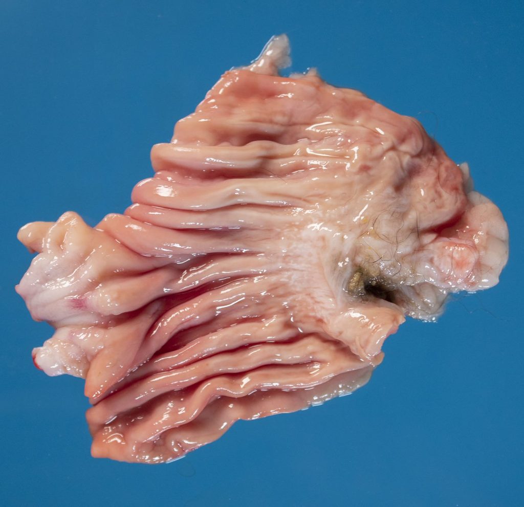
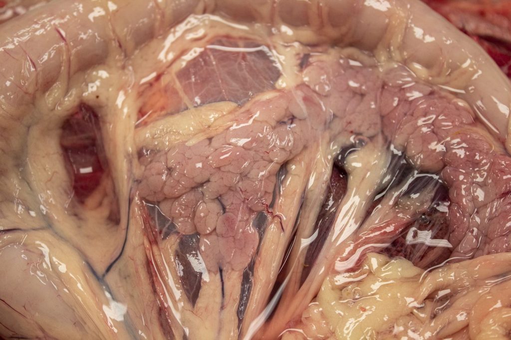
Above: Normal small intestine and mesen- tery, cat. The intestine is opened at left and the mucosa is visible. Notice that the small intestinal wall of carnivores is normally much thicker than that of herbivores.
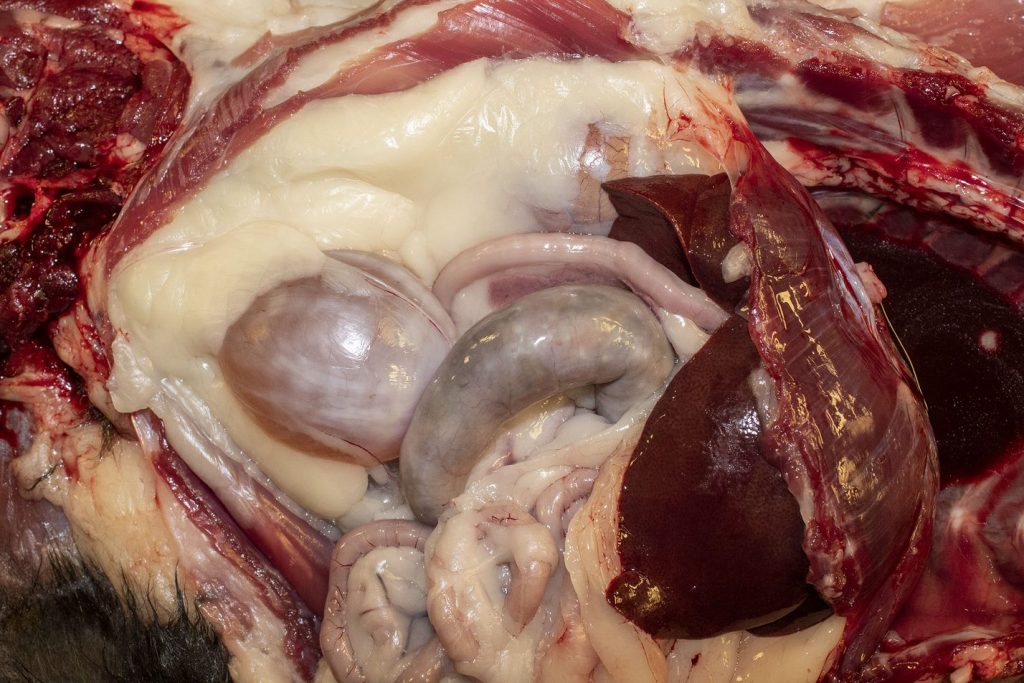
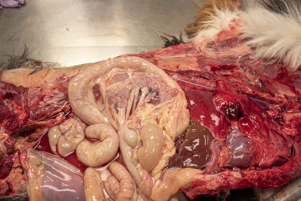
Above: Abdominal viscera, cat. Visible in the top photo are the diaphragm (right), liver, small intestine (centre: dorsal is duodenum, jejunum is at the bottom of the photo), colon (centre middle), and urinary bladder. The pancreas is visible in the mesentery of the duodenum (dorsal to the colon in the image). Note the abundant fat stores–the right kidney is peeking through the fat, dorsocaudal to the liver.
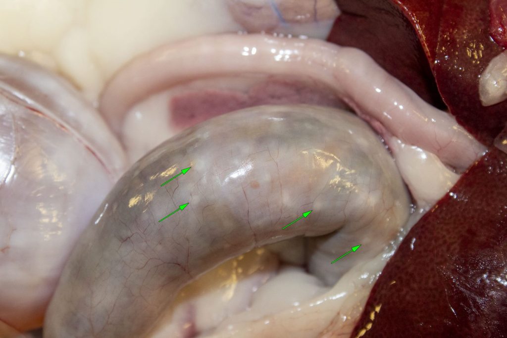
In the lower image, white-tan lymphoid nodules are visible in the colon (arrows). These are normal in young animals.
Above: Normal liver, cat.

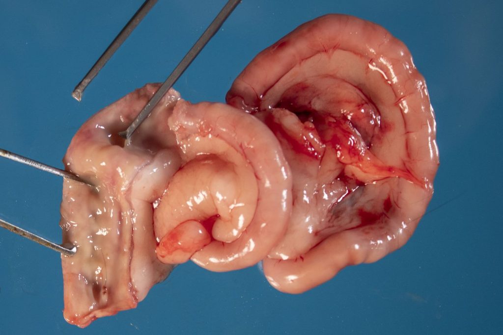
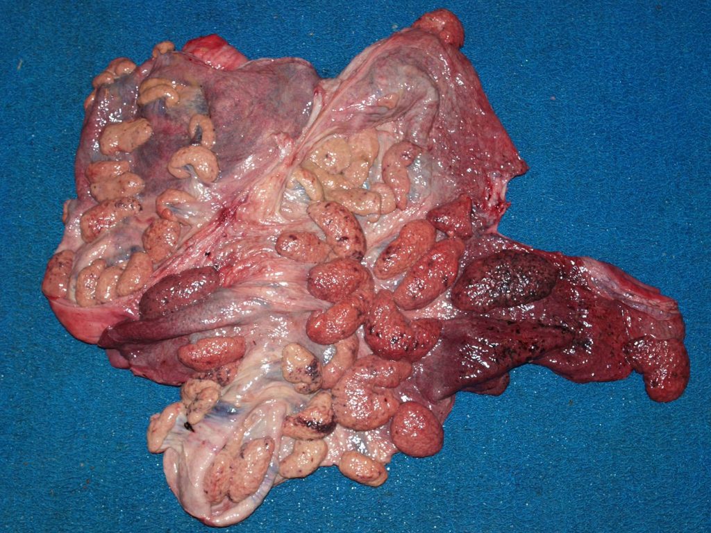
Above: Normal pancreas (and duodenum and mesentery), dog.
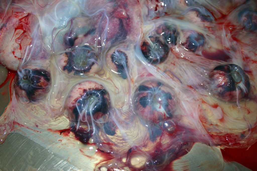
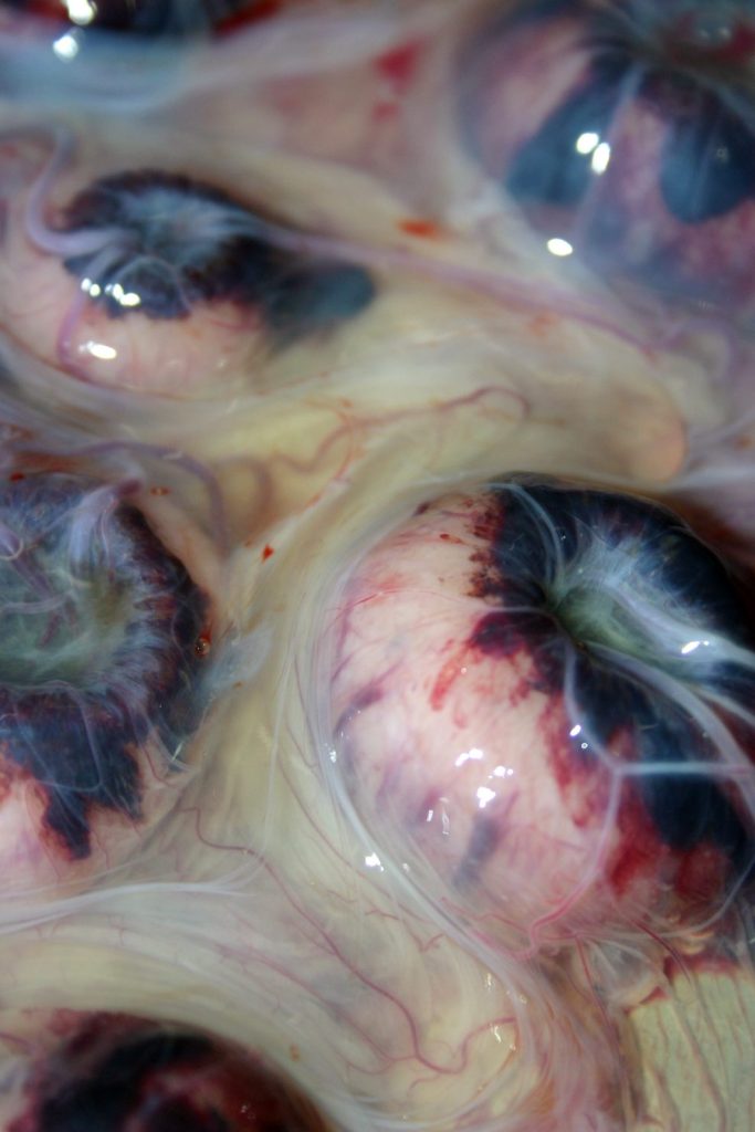
Above: Normal postpartum uterus, cow. The endometrial surface is shown, and cotyledons are visible.
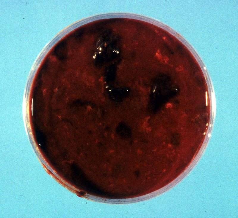
Above: Normal lochia (postpartum uterine fluid), cow. The lochia is opaque and red- black, but does not have a foul odour (in contrast to a uterus with bacterial infection).
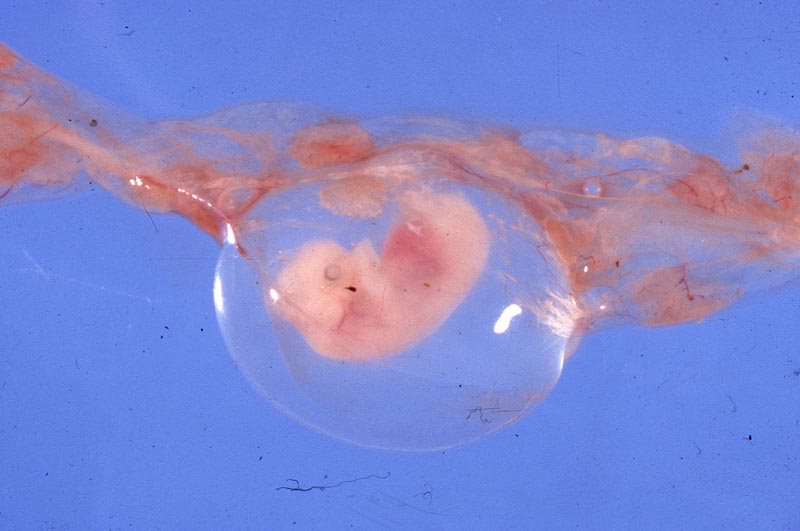
Above: Fetal lamb. The fetus is within the clear sphere of the amnion. The chorioalantois (with cotyledons) are visible at right, top and left; it would normally surround the amnion (the two membranes are separated by the allantoic cavity), and the chorioallantois would interface with the endometrium of the uterus.
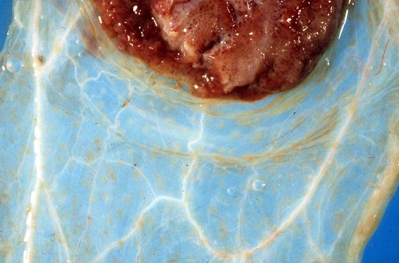
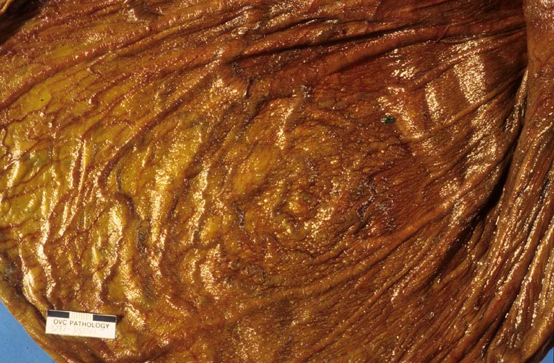
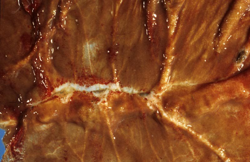
Above: Normal chorioallantois, bovine. A cotyledon is at the top. Note the thin transparency of the intercotylendonary area.
Above: Normal endometrium and placenta, sheep. The yellow-tan endometrium is covered
by a think tranparent chorioallantois. The white-pink caruncles (uterus) are more button-shaped than in cows, and are covered by a red cotyledon (placenta). Note the blood white blood vessels arising from the centre of the cotyledon, which will travel via the umbilical cord to the fetal lamb.
Above: Normal chorioallantois, horse. The chorionic surface of the equine chorioallantois has a diffusely villous or velvety appearance, except for a few non-villous areas as shown in the lower photo.
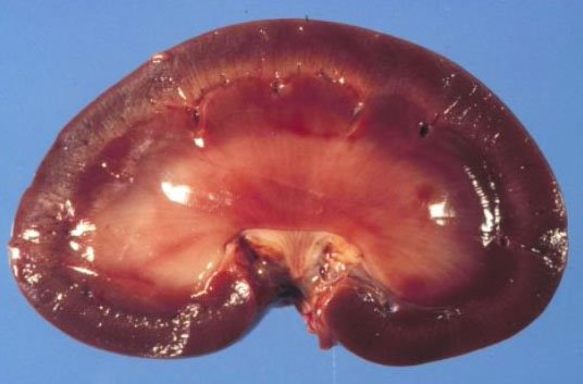
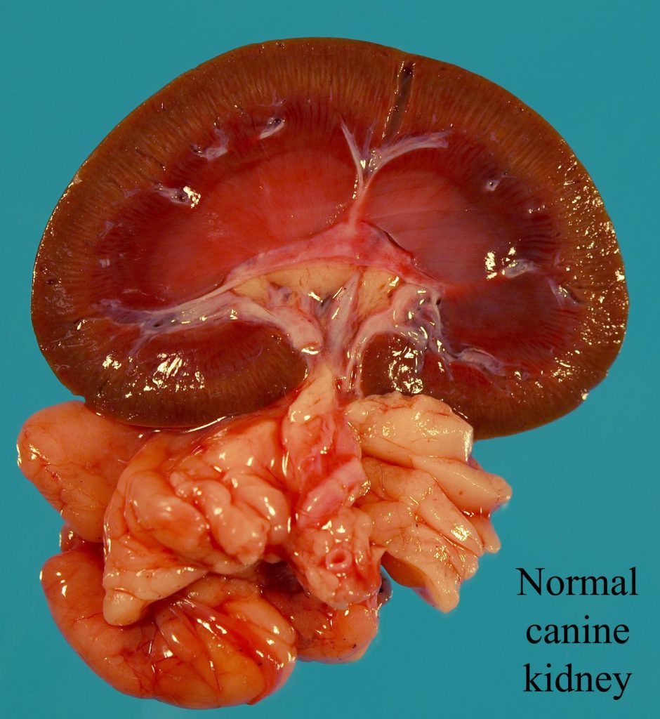
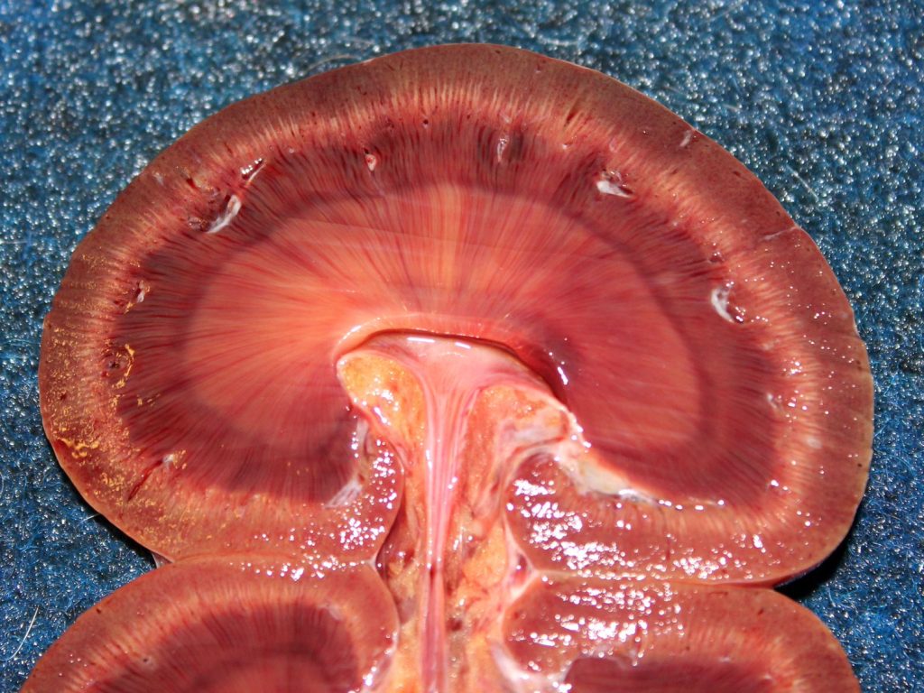
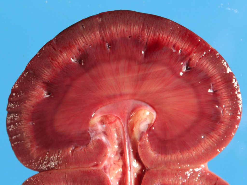
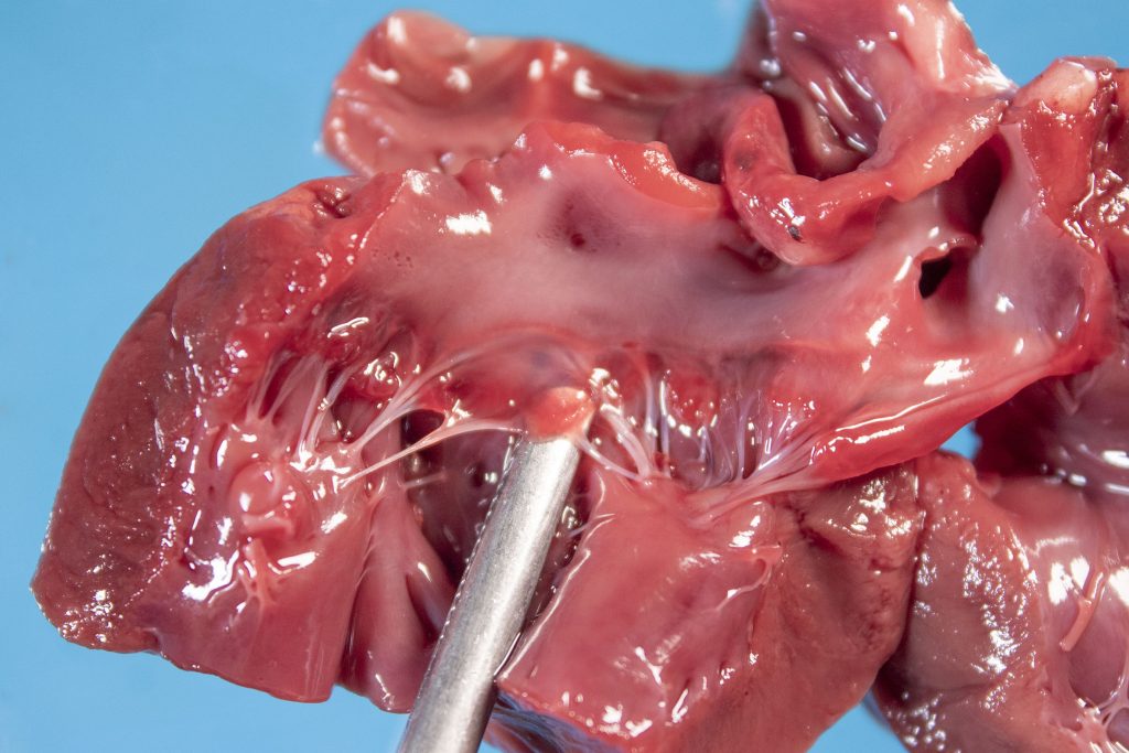
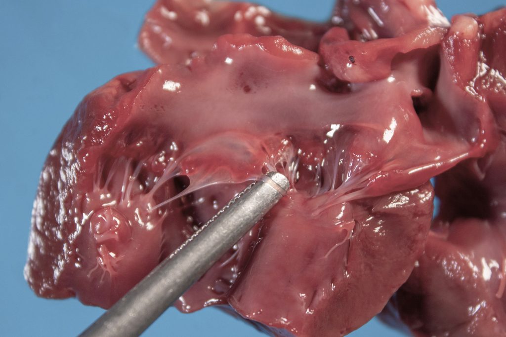
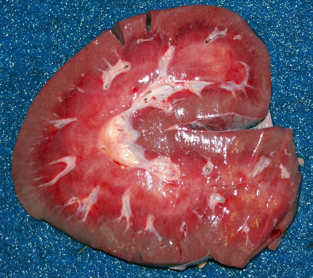
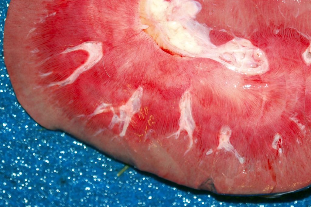
Above: Normal kidney, dogs. Note the capsular surface (top), cortex, corticomedullary junction containing arcuate arteries (visible in all of these photos), medulla, and pelvis. Note the pale fatty connective tissue around to the pelvis. Dog kidneys sometimes have peculiar morphologic features:
- The deep cortex is often pale. In the images in the centre column, radial stripes representing collecting ducts are visible within this arc of pallor.
- The outer medulla is often redder than the pale inner medulla.

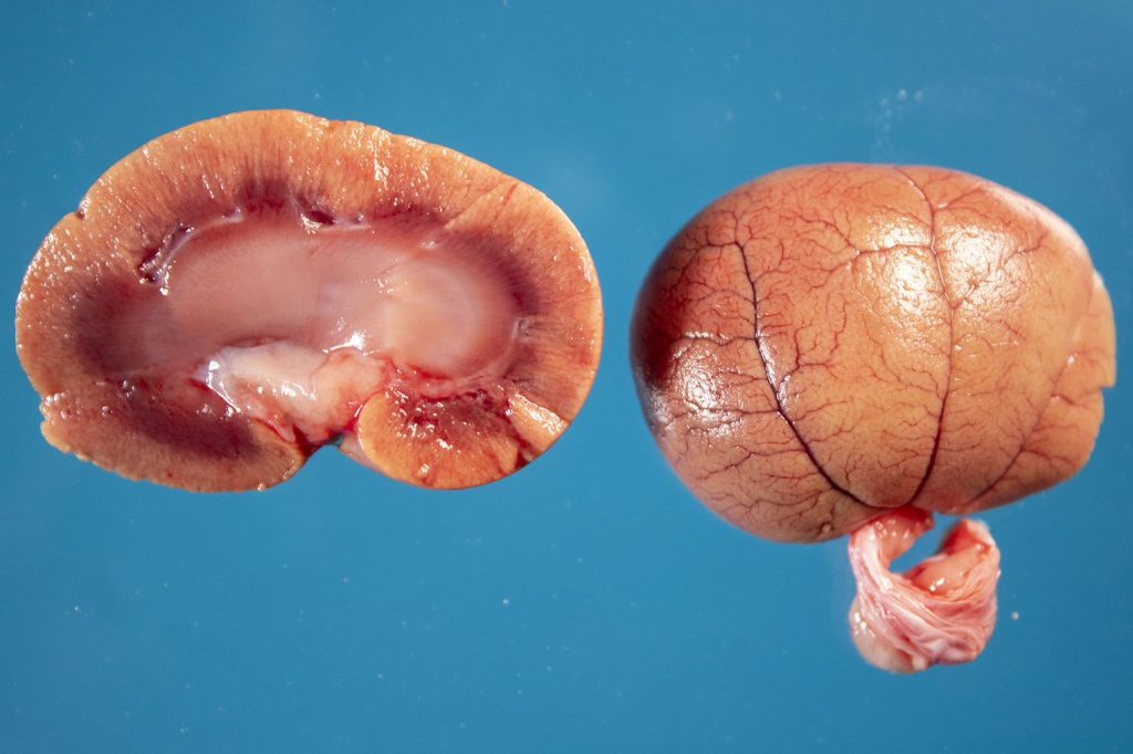
Above: Normal kidney, cat. Cat kidneys have a unique morphologic appearance:
- Blood vessels are prominently visible on the capsular suface
- The cortex is usually yellow, because of fat droplets within the cytoplasm of the proximal convoluted tubules.
Above: Normal kidney, horse. Although not visible in these photos, the pelvis and urinary bladder of horses often contains pasty white crystalline material because of the high calcium carbonate content of horse urine.
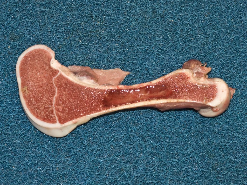
Above: Normal humerus, calf. Proximal at right. In the distal femur, not the white line of the physis, that separates the epiphysis (left) from the metaphysis (right of the physis). Note the following:
- The cortices
- Cancellous bone filling the epiphysis and metaphysis
- Tan-red fat with an absence of bone, in the marrow cavity of the diaphysis (centre).
Above: Normal distal limb, cow. Note the following:
- Dorsal surface of P3 is parallel to the dorsal hoof wall; these are separated by the laminae, and bound dorsally by the coronary band
- P1, P2 and P3 pedal bones, proximal and distal (navicular) sesamoid bones
- Distal piece of the metacarpal/metatarsal bone at right of upper image
- Joints: fetlock (at right of upper image), P1-P2 (pastern joint), P2-P3 (coffin joint)
- Deep digital flexor tendon
