Week 4: Dental Charting
Welcome to week 4! This week you will spend some time reviewing earlier concepts about the teeth including surfaces and labelling, and learn about the dental chart.
Review
Dental charting combines information from week 2 (tooth labelling) and week 3 (dental conditions and procedures).
Prior to moving forward, go back to weeks 2 & 3 in your course pack to complete the review exercises. This will ensure you have a strong foundation heading into dental charting!
Key Components of the Dental Chart
- Patient registration forms (medical and dental history, consent forms)
- Diagnosis and treatment plan documents
- Radiographs (x-rays)
- Financial information including private insurance
- Odontograms (chart used to indicate a client’s existing and planned dental conditions and procedures)
Sample Dental Chart
This screen capture of a dental chart on Wikipedia Commons [opens new tab] includes several of the elements outlined above.
Dental Records
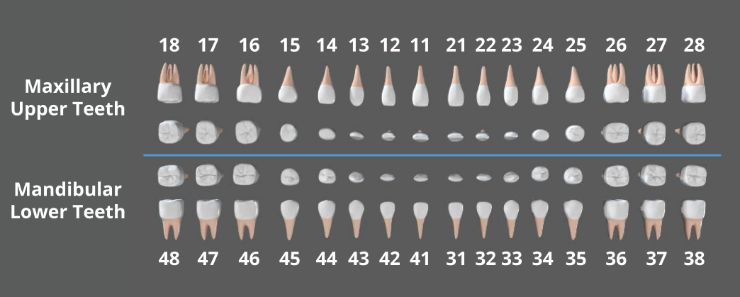
Odontogram Views
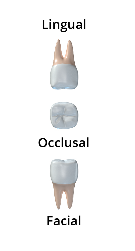
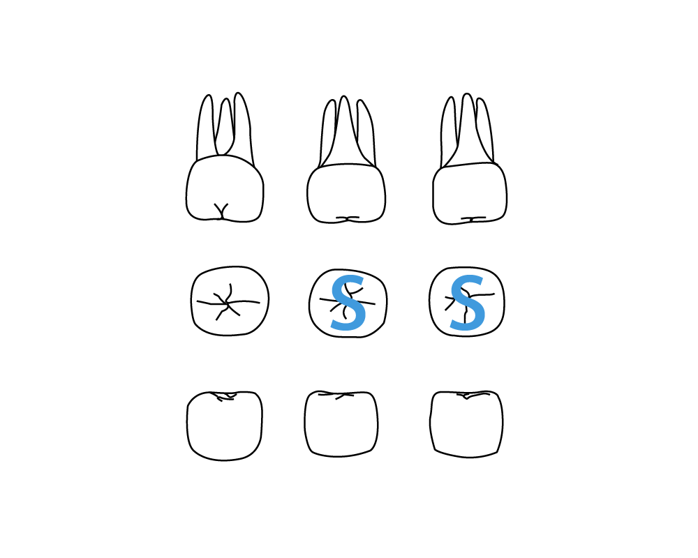
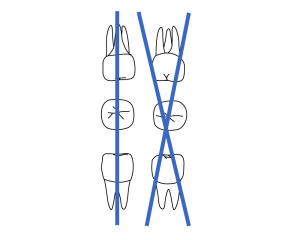
There are 3 primary views on odontograms
- Facial/buccal: how the tooth appears at the front
- Occlusal/incisal: how the tooth appears from the biting surface
- Lingual: how the tooth appears from the tongue
Sample Odontogram
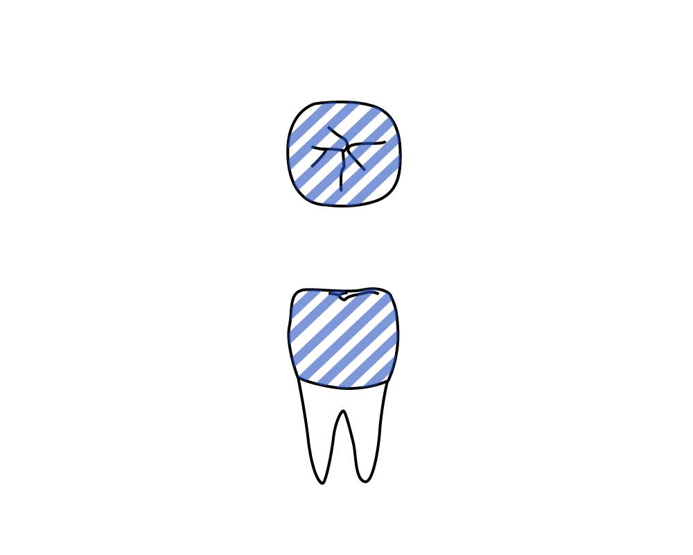
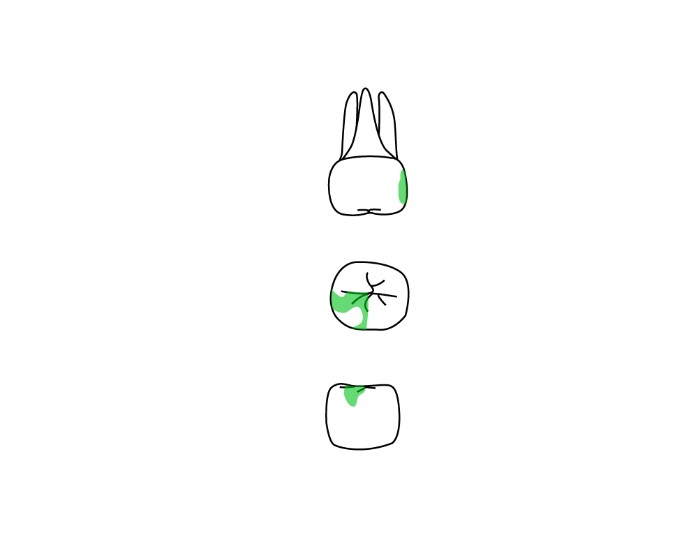
Charting Symbols
| Symbol: | Description: | Abbreviation: | Meaning: |
|
Blue shading | Am | Amalgam filling present |
|
Tan/green shading | Cr | Composite resin filling present |
|
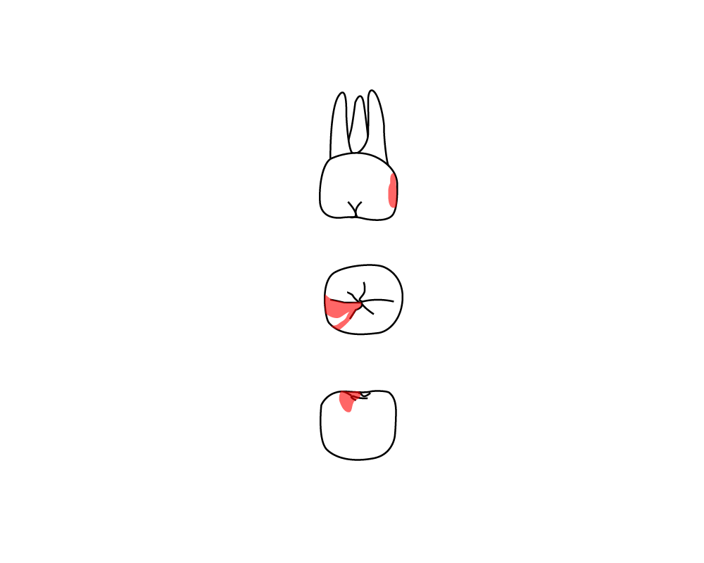
Red shading | n/a | Caries – tooth needs future filling |
|
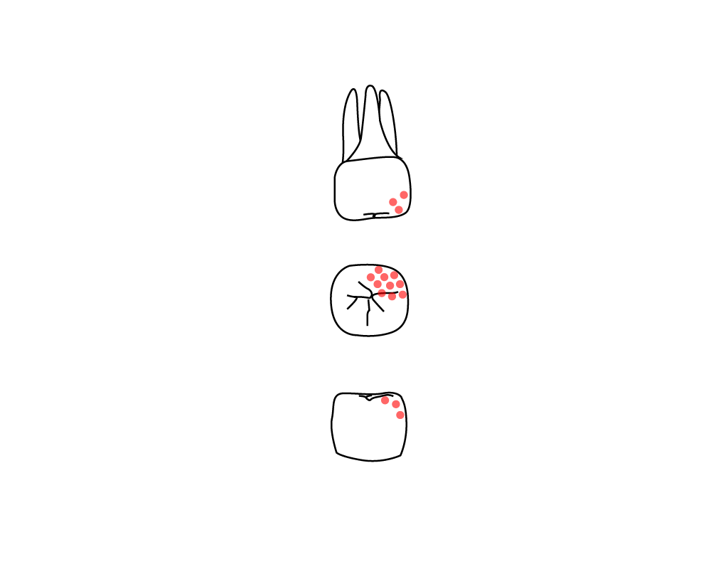
Red dots | incip | Incipient caries (early decay) |
|
n/a
|
S | Dental sealant was applied |
|
An X through the whole tooth | n/a | Tooth is missing/has been extracted |
|
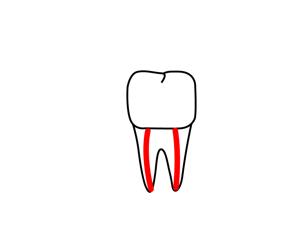
2 red lines through the root | RCT | Root canal therapy |
|
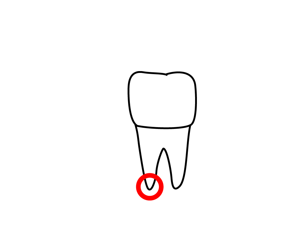
Small red circle at the base of the root | n/a | Abscess |
|
Multiple teeth connected with a line | n/a | A bridge |
|
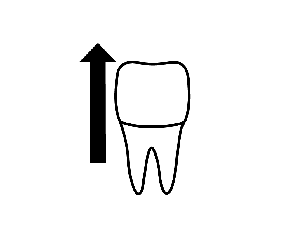
An arrow pointing towards the crown of the tooth | n/a | An over-erupted tooth |
|
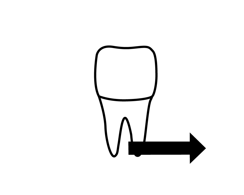
An arrow pointing away from the midline of the tooth | n/a | Shows the direction of a tooth which is drifting |
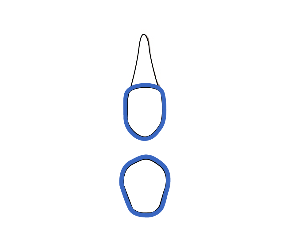 |
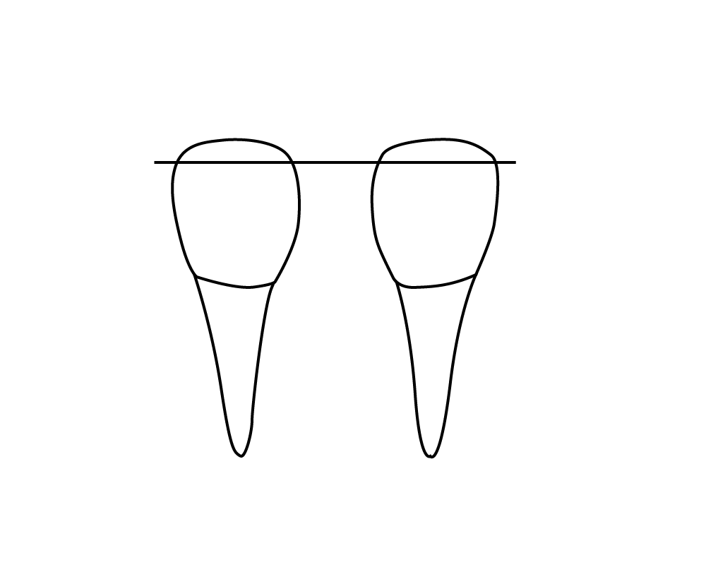
Blue outline of the facial surface of the tooth | n/a | Veneer present |
References
Torres, H. O., Ehrlich, A., Bird, D. & Dietz, E. (2009). Modern dental assisting (9th ed.). W.B. Saunders Company.
Baillargeon, S. (2008). Dental office administration. Thomson Nelson.
