Week 2: Tooth Structure and Identification
Welcome to week 2! During class you will be become familiar with the anatomy of the mouth and teeth and learn about Canada’s preferred dental tooth numbering system. This information will serve as an important foundation for future topics in this course such as dental conditions and procedures, charting, and insurance. There are several great knowledge checks available to you this week, so be sure to complete all the activities!
Dental Arches
The maxillary arch (upper arch) is part of the skull, and not capable of movement. The mandibular arch (lower arch) is moveable through the temporomandibular joint (TMJ). Occlusion refers to when teeth of both arches are in contact.
Dental Quadrants
The 2 arches are divided into 4 equal sections called quadrants.
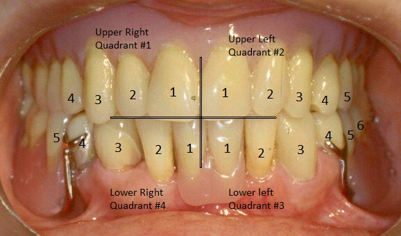
Use your knowledge of the upper and lower arch names to fill in the correct dental quadrant name below:
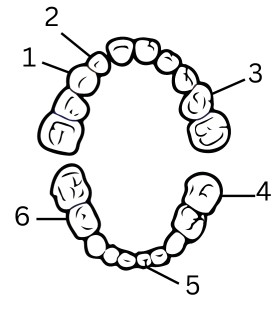
Dental Sextants
The teeth are also divided into 6 equal parts called sextants. This includes upper front, left/right, and lower front, left/right.
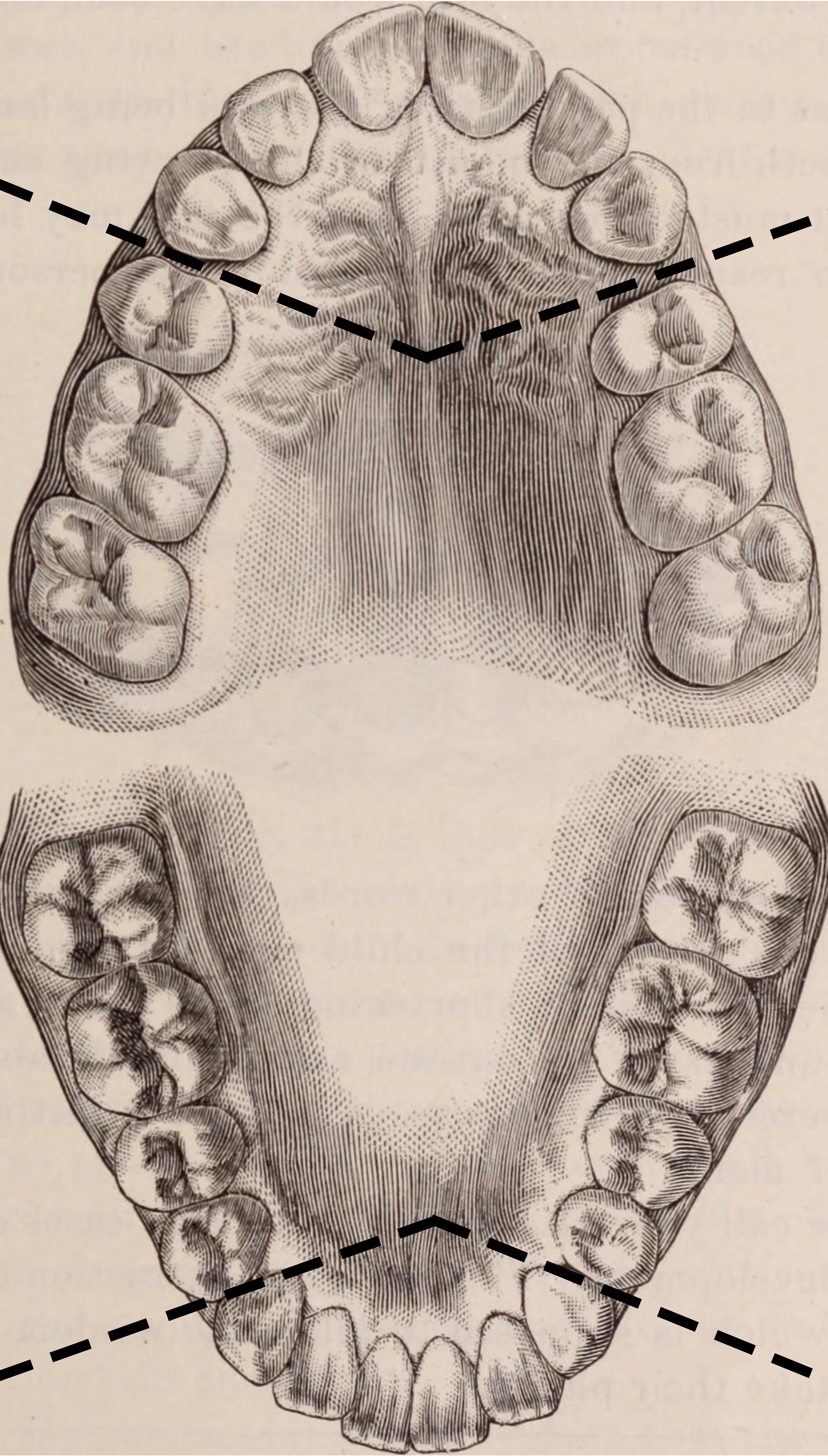
Use your knowledge of the upper and lower arch names to fill in the correct dental sextant names below:
Types of Teeth
The 4 Types of Teeth:
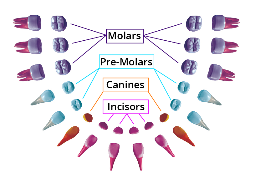
Incisors (Pink): Made up of central incisors and lateral incisors. Single rooted with a thin edge.
Cuspids/Canines (Orange): The longest and most stable teeth with the longest roots.
Bicuspids/Premolars (Blue): Have a broad chewing surface designed to grind food.
Molars (Purple): Shorter and blunt with the largest surface for chewing.
Primary Vs. Permanent Dentition
Primary dentition is also known as deciduous dentition or baby teeth. These are the first set of 20 teeth that are usually present by age 2. The mandibular central incisors are among the first teeth to erupt, with mandibular molars among the last. Primary teeth can decay more quickly because the enamel and dentin are thinner. This means that good oral hygiene practices are even more important among children.
Mixed dentition refers to the period of time where children have a mixture of both permanent and primary teeth. Crowding may begin as the jawbones begin to grow accommodate larger permanent teeth. This can cause pain, discomfort, and embarrassment due to differences among size and spacing.
Permanent dentition is also known as adult teeth. On average this consists of 32 teeth and fully appear once the last primary tooth is shed by age 14. The except is 3rd molars which usually appear by age 18.
International Tooth Identification System
The International (FDI) Tooth Identification System is the most commonly used dental labelling system in Canada. The Universal Numbering System is commonly used in the United States. The Palmer Notation System is commonly used in the United Kingdom.
We will focus our learning material on the international (FDI) system.
Tooth labelling will come up again when we explore dental insurance!
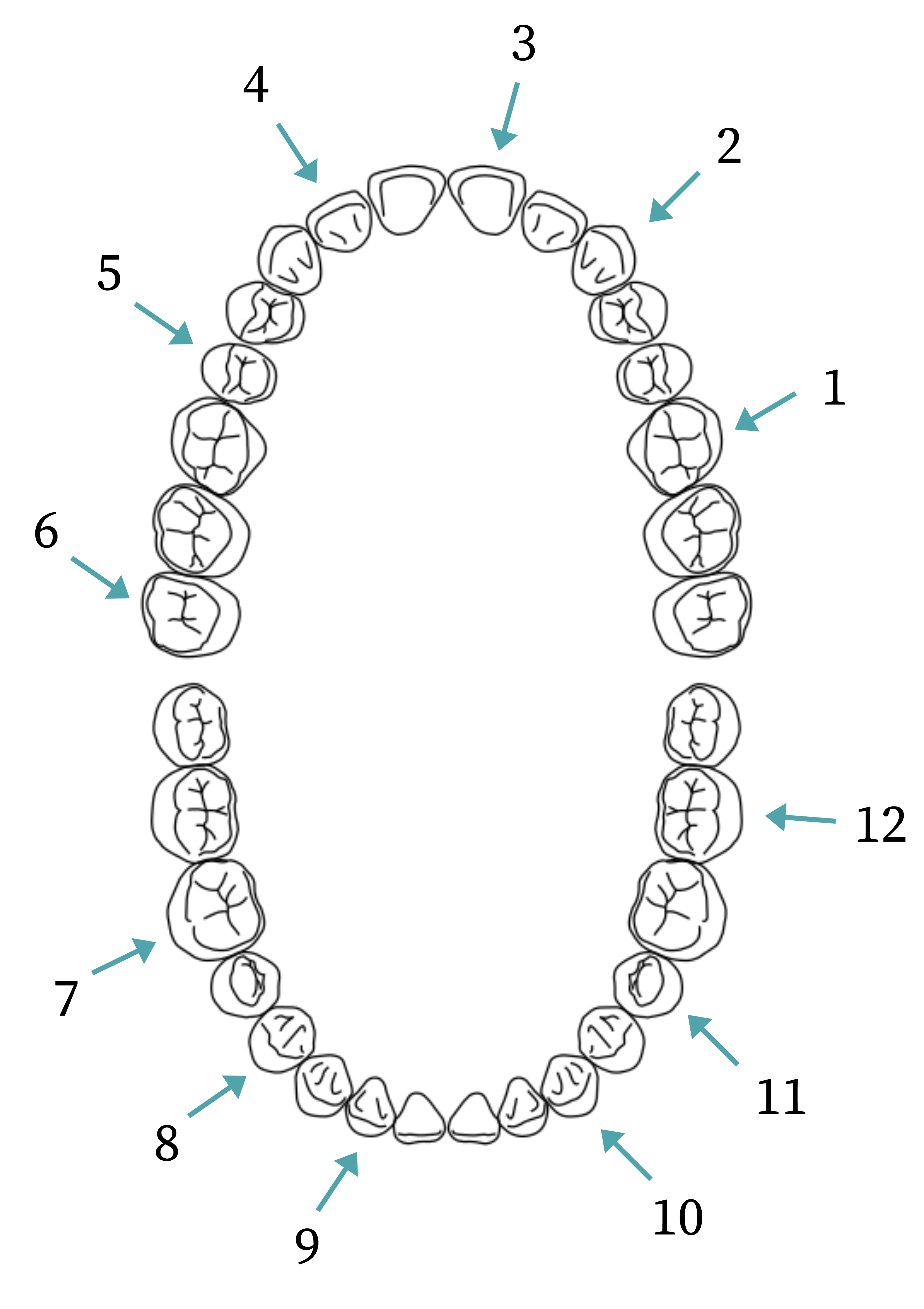
Permanent Teeth and the International Tooth Identification System
Identify the permanent teeth using the International Tooth Identification System.
Primary Teeth and the International Tooth Identification System
Identify the primary teeth using the International Tooth Identification System.
Tooth Surfaces
Each tooth has 5 surfaces.
Distal: the surface of the tooth further from the midline.
Mesial: the surface of the tooth closest to the midline.
Incisal (anterior) / Occlusal (posterior): the biting/chewing surface of the tooth.
Labial (anterior) / Facial or vestibular (posterior): the surface facing the inside of the face/cheek/gums.
Lingual / palatal: the surface of the tooth closest to the tongue
Tooth surfaces will come up again when we explore dental insurance!
Tooth Anatomy
Review the key components of tooth anatomy below:
References
Torres, H. O., Ehrlich, A., Bird, D. & Dietz, E. (2009). Modern dental assisting (9th ed.). W.B. Saunders Company.
Baillargeon, S. (2008). Dental office administration. Thomson Nelson.
