9.2
The three different types of dental x-ray film are: intraoral film, extraoral film, and duplicating film.
Intraoral Film
The intraoral film is placed inside the mouth and used to examine the teeth and supporting structures. The terms “film” and “film packet” are often used interchangeably. The dental film serves as a recording medium and is used in traditional film radiography, and a medium in digital radiography is known as a receptor.
Essential questions to ask yourself when dealing with intraoral film:
- How do skilled dental radiographers reduce the patient’s exposure to x-rays? – By reducing the number of retakes.
- Why does the film need to be protected from the light? – Light is a form of radiation and will expose and ruin the film. Light cannot pass through the packet, but x-rays can.
Intraoral Film Packaging
The packaging for intraoral film is used to protect the film from light and moisture. It is usually available in plastic trays or cardboard boxes containing 25, 100, or 150 films and the boxes are labeled with the type of film, film speed, film size, number of films per packet, total number of films enclosed, and the expiration date. Some packets contain two films and this allows for a copy to be made instantly when it is known that a copy will be needed, such as with certain insurance claims or specialty referrals. It also eliminates the need to duplicate.
In the Iannucci & Howerton, Dental Radiography Principles & Techniques, 6th Edition textbook on page 80, refer to Figure 9-4.
Film packets protect the film from light and moisture and have four components: x-ray film, paper film wrapper, lead foil sheet, and outer package wrapping.
In the Iannucci & Howerton, Dental Radiography Principles & Techniques, 6th Edition textbook on page 80, refer to Figure 9-5.
Intraoral Film Types
There are three intraoral film types: periapical film, bite-wing film, and occlusal film.
Periapical Film
The periapical film is used to examine the entire tooth and supporting bone. This type of film shows the tip of the tooth root and surrounding structures, as well as the crown.
In the Iannucci & Howerton, Dental Radiography Principles & Techniques, 6th Edition textbook on page 82, refer to Figure 9-9.
Here is an image of a dental periapical x-ray showing the details of the lower molars and surrounding bone structure.
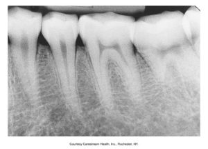
Periapical films come in different sizes.
| Sizes | Used For |
| Size 0 | Used for small children |
| Size 1 | Used for anterior teeth in adults |
| Size 2 | Standard film, used for anterior and posterior teeth in adults |
Size #2 film is typically used for both anterior and posterior views because it will give a wider image that will overlap adjacent images. This allows for areas to be represented more than once in a set of x-rays, acting as a diagnostic backup. This reduces the need for retakes without increasing the amount of radiation exposure for the patient.
Bite-Wing Film
Bite-wing film is used to examine the crowns of both maxillary and mandibular teeth on one film and for examining interproximal surfaces. Stick-on tabs of bite-wing loops. can be used, and the bite-wing film has a “wing,” or tab, attached to the tube side of the film. Bite-wing films are helpful in detecting cavities in between the posterior teeth.
In the Iannucci & Howerton, Dental Radiography Principles & Techniques, 6th Edition textbook on page 82, refer to Figures 9-10 & 9-11.
Here is an image of a dental bite-wing x-ray displaying the upper and lower posterior teeth and another image of what a bite-wing tab holder looks like.
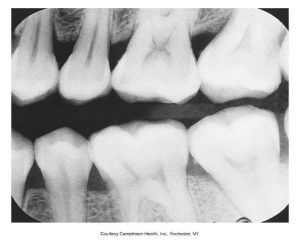
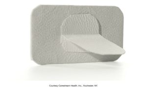
Bite-wing films come in different sizes.
| Sizes | Used For |
| Size 0 | Used for posterior teeth in small children |
| Size 2 | Used for posterior teeth in older children or adults |
| Size 3 | Longer and narrower than standard size 2; used only for bite-wing for adults |
Size #3 is currently not recommended because not all the contacts can be opened on one film.
Occlusal Film
The occlusal film is a larger film used for examination of large areas of the maxilla or mandible. This film was named occlusal because the patient “occludes,” or bites on, the entire film.
In the Iannucci & Howerton, Dental Radiography Principles & Techniques, 6th Edition textbook on page 83, refer to Figure 9-12.
Here is an image of an occlusal dental x-ray showing a full arch view of the upper or lower teeth.
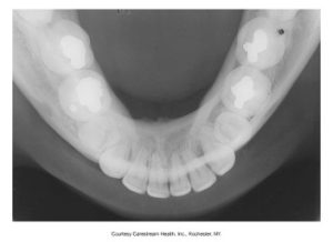
There is a size #4 occlusal film that is used to examine large areas of the maxilla or mandible, and it is almost four times the size of standard #2 film.
Intraoral Film Sizes
Intraoral film sizes range from 0 to 4 the different types are periapical film, bite-wing film, and occlusal film, which you just covered in this chapter.
In the Iannucci & Howerton, Dental Radiography Principles & Techniques, 6th Edition textbook on page 83, refer to Figure 9-13.
Below is a chart of the different sizes of intraoral dental X-ray films, ranging from Size 0 to Size 4, each with dimensions provided in millimeters and inches.

Intraoral Film Speed
Film speed is the amount of radiation required to produce a dental image of standard density that is determined by the size of silver halide crystals and the thickness of the emulsion. Larger crystals increase film speed but slightly reduce image resolution and the presence of special radiosensitive dyes. A fast film requires less radiation exposure.
X-ray films are given speed ratings ranging from A (slowest) to F (fastest). Only D-speed, E-speed, E/F-speed, and F-speed are used for intraoral radiography. The use of F-speed film results in less radiation exposure for the patient. F-speed films reduce radiation dose by 60% to the patient but also provide stable contrast characteristics under various processing conditions. Film speed can be found on the label side of the intraoral film packet, as well as on the outside of the film box.
Extraoral Film
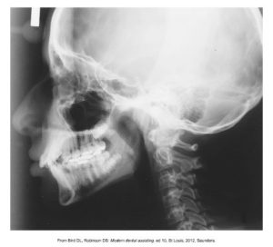
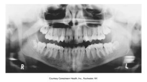
The extraoral film is placed outside the mouth during exposure and is used to examine large areas of teeth and jaws. The panoramic film is for a wide view of the upper and lower jaws and the cephalometric film is for bony and soft tissue areas in profile (from the side).
Extraoral films are more common in specialty practices, such as orthodontic, pediatric, and oral surgery practices.
Orthodontists commonly use cephalometric films to view the relationship of the jaw to the skull.
Below is an image of a cephalometric radiograph showing the profile of the cranial bone structure, jaw, and teeth.
Pediatric dentists use panoramic films to see the developing teeth because it is difficult for a child to sit through a full-mouth series of dental images. It also exposes the child to less radiation than a full-mouth series.
Below is an image of a panoramic dental x-ray image showing a full view of the upper and lower teeth and jaws, marked with “R” for the patient’s right side and “L” for the left side.
In the Iannucci & Howerton, Dental Radiography Principles & Techniques, 6th Edition textbook on page 84, refer to Figures 9-14 & 9-15.
Extraoral Film Packaging
Extraoral film packaging is boxed in quantities of 50 or 100 films and is not enclosed in moisture-proof packs. Extraoral films should be stored in a darkroom as this will prevent them from being accidentally opened in a lighted room, which can expose and ruin all the film in the box. The sizes of extraoral film packaging is 5×7 inch or 8×10 inch sizes, and 5×12 inch or 6×12 inch sizes for panoramic films.
In the Iannucci & Howerton, Dental Radiography Principles & Techniques, 6th Edition textbook on page 84, refer to Figure 9-16.
Media Attributions
- Iannucci & Howerton: Dental Radiography Princip, 6th Edition, Chapter 9, CC BY-NC-ND

