5.2
Amperage and Milliamperage
Amperage determines the amount of electrons passing through the cathode filament. Increasing amperage results in an increased number of electrons traveling from cathode to anode and the production of an increased number of x-rays. The quantity of the x-rays produced is controlled by milliamperage.
| Ampere (A) |
|
| Milliampere (mA) |
|
Milliamperage regulates the temperature of the cathode filament. A higher milliampere setting increases the temperature of the cathode filament and it increases the number of electrons produced and it also increases the number of x-rays emitted from the tube.
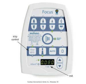
Here is an image of a control panel of a dental X-ray unit displaying buttons for various functions and indicators, including kilovoltage (kVp) and milliamperage (mA) settings.
Milliampere-seconds
mAs is the product of milliamperes and exposure time. When milliamperage is increased, the exposure time must be decreased to maintain a constant density.
Density and Milliamperage
An increase in milliamperage increases the overall density of an image, which results in a darker image.
Exposure time and Milliamperage
An inverse relationship is when milliamperage is increased, and the exposure time must be decreased. When milliamperage is decreased, the exposure time must be increased.
Exposure Factor Tips
All dental x-ray machines have three exposure factor settings: kV, mA, and time.
X-ray Beam Intensity
The key factors of x-ray beam intensity are kilovoltage peak, milliamperage, exposure time, distance, inverse square law, and half-value layer. The product of the quantity (number of x-ray photons) and quality (energy of each photon) per unit of area per unit of time of exposure.
Media Attributions
- Iannucci & Howerton: Dental Radiography Principles and Techniques, 6th Edition, Chapter 5, CC BY-NC-ND
