4.3
Operator Protection
Dental radiographers must accept responsibility for their own radiation safety, protection guidelines, and radiation monitoring.
Protection Guidelines
The dental radiographer must avoid the primary beam. Protection guidelines include distance recommendations, position recommendations, and shielding recommendations. To keep radiation exposure to zero, the dental radiographer must carefully follow the safety guidelines and use radiation monitoring devices.
Distance Recommendations
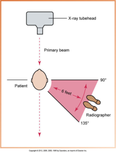
Maintain an adequate distance during exposure. The dental radiographer must maintain a distance of at least 6 feet from the tubehead during exposure. When maintaining this distance is not possible, a protective barrier must be used. Avoid the primary beam by standing either perpendicular or at a 90- to 135-degree angle to the beam. To avoid the primary beam, proper operator position during exposure includes the dental radiographer never holding a receptor in place for a patient, and the dental radiographer must never be holding or stabilizing the x-ray tubehead.
Safety Code 30 – Protection of the operator and others near dental X-ray equipment should be achieved by:
- Ensuring that the room containing the dental X-ray equipment is designed so that during the examination, the operator is not exposed to the primary radiation beam and can keep a distance of at least 3 meters from the X-ray tube and from the patient.
- If it is not possible for an operator to keep at a distance of at least 3 meters (9 feet) from the X-ray tube, an adequately shielded barrier, which allows observation of the patient, must be provided for the operator to stand behind during radiography.
Operator Protection Guidelines
Operator protection guidelines suggest that the dental radiographer stand at an angle of 90 to 135 degrees to the primary beam.
The diagram below shows operator protection guidelines during an x-ray procedure with the x-ray tubehead. The direction of the primary beam and the recommended position and angle for the radiographer to stand at a safe distance from radiation.
Shielding Recommendations
Dental office design may include walls to protect the operator from primary and scatter radiation. There may be a lead wall to stand behind, or you may need to walk around the corner with a portable exposure button. There may be an exposure button positioned on a wall outside the room used for radiographs.
Safety Code 30 – General Information on Shielding Tables:
- The following information applies to shielding tables in this Appendix. 1.
- The tabulated values give the minimum amount of lead, gypsum, and concrete shielding required to reduce the skin dose in uncontrolled areas to 1 mSv in one year and in controlled areas to 20 mSv in one year.
Radiation Monitoring
Equipment monitoring is when dental x-ray machines must be monitored for leakage radiation, and personnel monitoring is a film badge (Dosimeter) that can be worn at waist level when taking radiographs. It is then mailed along with a control badge to the monitoring company once a month for evaluation.

Radiation Exposure Guidelines
The radiation exposure guidelines are:
- Radiation Safety Legislation
- Maximum Permissible Dose
- Maximum Accumulated Dose
- Cumulative Occupational Dose
- ALARA Concept
- Radiation exposure guidelines have been established to protect the patient and operator from excess radiation.
- Strict adherence to radiation exposure guidelines is mandatory for all dental radiographers.
Please refer to your textbook to see Table 4-1 Pgs.34-35 Recommendations for Prescribing Dental Radiographs (2022)
Radiation Safety Legislation
The dental radiographer must be familiar with the laws that apply to their workplace and jurisdiction.
- Healing Arts Radiation Protection (HARP) Act: Provincial regulation of x-rays in Ontario
- Safety Code 30: Radiation Protection in Dentistry in Canada
Maximum Permissible Dose (MPD)
MPD is the maximum dose equivalent that a body is permitted to receive in a specific period.
- MPD for occupationally exposed persons is 5.0 rem/year,
- For nonoccupationally exposed persons, it is 0.1 rem/year.
- For occupationally exposed pregnant women, MPD is the same as for a nonoccupationally exposed person – 0.1 rem/year.
Dental personnel should strive for an occupational dose of 0 by adhering to strict radiation protection practices.
Maximum Accumulated Dose (MAD)
- MAD is the dose accumulated over a lifetime. A formula based on the worker’s age:
- MAD = (N – 18) X 5 rem/year
- MAD = (N – 18) X 0.05 Sv/year
And the age of 18 is the minimum required age for a person who works with radiation.
Cumulative Occupational Dose
Cumulative occupational dose is the dose accumulated over a lifetime. An individual’s cumulative occupational effective dose should not exceed the worker’s age multiplied by 10 mSv.
ALARA Concept
ALARA concept means that the level should be as low as reasonably achievable, and every possible method of reducing exposure to radiation should be employed.
Some precautions taken to abide by the ALARA concept are:
- Radiographs should be ordered only for diagnostic purposes
- Use the lowest possible kVp, mA, and exposure time use F-speed film or digital radiography
- Use a longer PID with a rectangular shape;
- Use a tubehead with an aluminum filter and a lead collimator
- Use a lead apron and thyroid collar
- Use film-holding devices
- Avoid retakes
- Test equipment for efficiency and proper functioning
Radiation Protection and Patient Education
Patient education is everything you say to the patient before, during, and after the procedure. It may be useful to have educational brochures on hand that explain the importance of diagnostic dental radiographs and how their benefits far outweigh the minimal risks. Some dental facilities also have informative videos that can help educate patients about dental radiographs. Patient education may take the form of an informal conversation or printed literature, and the dental radiographer must be prepared to explain exactly how patients are protected before, during, and after x-ray exposure.
Media Attributions
- Iannucci & Howerton: Dental Radiography Principles and Technique, 6th Edition, Chapter 4,CC BY-NC-ND
