4.2
During Exposure
As a dental radiographer, you want to limit the amount of radiation received by the patient. Proper selection of exposure factors and good technique further protect the patient from excess radiation exposure.
Exposure factors are:
- Thyroid collar
- Lead apron
- Image Receptors
- Beam Alignment devices
- Exposure factor selection
- Proper technique
Thyroid Collar
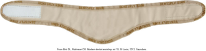
A thyroid collar is a flexible lead shield placed around the patient’s neck that protects the thyroid gland from scatter radiation. A thyroid collar may be separate or part of a lead apron. It is recommended that the patient wear it for all intraoral exposures and not for extraoral exposures. Below is an image of what a typical thyroid collar looks like.
Lead Apron
A lead apron is placed over the patient’s chest and lap to protect the reproductive organs and blood-forming tissues from scatter radiation and the use of lead aprons are often a state/provincial law.
For Safety Code 30 (Section 9.2):
- The patient must be provided with a shielded apron for gonad protection and a thyroid shield, especially during occlusal radiographic examinations of the maxilla.
- The use of a thyroid shield is especially important in children.
- The shielded apron and thyroid shield should have a lead equivalence of at least 0.25 mm of lead.
- In panoramic radiography, since the radiation also comes from the back of the patient, a conventional lead apron is not adequate, and dual (front and back) lead aprons should be worn.
Dental radiographers should ensure that the lead apron and thyroid collar are stored properly and not folded since creases may crack the lead. Care should also be taken not to touch the apron with the same gloves that were used to place the film in the patient’s mouth, as this will cause cross-contamination, compromising infection control. The radiographer should ensure that the patient removes any oral piercings, eyeglasses, earrings, and removable dental prostheses that may distort or interfere with the desired image.
The image below displays examples of lead aprons. As mentioned, a thyroid collar may be attached to the lead apron or used as a separate shield.

Image Receptors
Digital image receptors require less radiation exposure for the patient, and the use of a digital receptor is the most effective method of reducing a patient’s exposure to radiation.
Beam Alignment Device
Beam alignment devices stabilize the receptor in the mouth and reduce the chance of movement. It eliminates the need for the patient to hold the receptor in position with a finger, reducing unnecessary exposure.
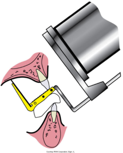
Below is an illustration of a dental x-ray beam alignment device, showing how it positions the film and the x-ray tube for an accurate exposure.
Exposure Factor Selection
Exposure factor selection is an adjustment of kVp, milliamperage, and time settings on a control panel to limit the amount of x-radiation exposure received by the patient. On most units, the kilovolt peak and milliamperage are preset by the manufacturer and cannot be adjusted. A setting of 70 to 90 kVp keeps patient exposure to a minimum.
Proper Technique
The proper technique for non-diagnostic images must be retaken, resulting in additional radiation exposure for the patient. Retakes are a major cause of unnecessary radiation to patients and must be avoided at all times. Having to retake radiographs wastes time and is not relished by most patients. If a retake is ordered, the dental radiographer should know how to correct the error that resulted in the need for a retake.
After Exposure
Proper receptor handling artifacts caused by improper receptor handling result in nondiagnostic images and careful handling is crucial.
Proper image retrieval is improper image retrieval, which may require retakes, needlessly exposing the patient to excess x-radiation.
Media Attributions
- Iannucci: & Howerton Dental Radiography Principles and Technique, 6th Edition, Chapter 4, CC BY-NC-ND
