33.2
Classification of Caries on Dental Images
The image appearance of dental caries can be classified according to the location of the caries on the tooth.
Interproximal Caries
Interproximal caries are found between two adjacent surfaces, and are typically seen on dental images at or just below the contact point. As caries progresses through the enamel, it typically assumes a triangular configuration. When it reaches the DEJ, it spreads laterally and progresses through dentin. These are classified as incipient, moderate, advanced, and severe.
Occlusal Caries
Occlusal caries are caries that involve the chewing surface of posterior teeth. Thorough clinical exam with a mouth mirror, explorer and light is the method of choice for the detection of occlusal caries. Early occlusal caries is difficult to see on a dental image.
Incipient Occlusal Caries
Incipient occlusal caries cannot be seen on a dental image, it must be detected with an explorer. Occlusal carious lesions can be classified as incipient, moderate, or severe.
Moderate Occlusal Caries
Moderate occlusal caries extends into dentin. It appears as a thin, radiolucent line located under the enamel of the occlusal surface of the tooth.
Severe Occlusal Caries
Severe occlusal caries extends into dentin and appears as a radiolucency. The radiolucency extends under the enamel of the occlusal surface of the tooth. Apparent clinically and appears as a cavitation in a tooth.
Buccal and Lingual Caries
These are difficult to detect on a dental image because they are superimposed on tooth structure. If seen on a dental image, they appear as a circular radiolucent area. To determine the location of the lesion, a clinical examination with an explorer is necessary.
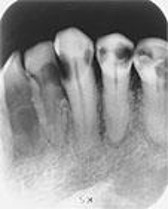
Root Surface Caries
Root surface caries involves only the roots of teeth. On a dental image, it appears as a cupped-out or crater-shaped radiolucency below the CEJ.
Early lesions may be difficult to detect on a dental image. Bone loss and corresponding gingival recession precede the caries process and result in exposed root surfaces. Most common locations include the exposed roots of the mandibular premolar and molar areas.

Root caries appearing as a crater shaped radiolucency just below the cemento-enamel junction (CEJ) on the mandibular 2nd premolar.
Recurrent Caries
Recurrent caries occurs adjacent to an existing restoration. It appears as a radiolucent area just beneath a restoration. It is most often located beneath the interproximal margins of a restoration. Occurs because of inadequate cavity preparation, defective margins, or incomplete removal of caries before place of the restoration.
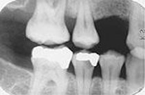
Recurrent caries seen as a radiolucency below a two-surface amalgam restoration on the mandibular second premolar.
Rampant Caries
Rampant caries are advanced and severe caries affecting several number of teeth. This is associated with children with poor diets and adults with decreased salivary flow.
Rampant Caries
Conditions Resembling Caries
On a dental image, conditions that may be confused with caries include:
- Cervical burnout
- Restorative materials
- Attrition
- Abrasion
Media Attributions
- Images courtesy of Iannucci & Howerton: Dental Radiography Principles & Techniques, 6th Edition, 2022, Chapter 33.
