32.1
Identification of Restorations
Metallic restorations |
Metallic restorations absorb x-rays. Very little radiation contacts the receptor. That area of receptor remains unexposed, and the metallic restorations appear completely radiopaque on a dental image. |
Nonmetallic restorations |
Nonmetallic restorations may vary in appearance from radiolucent to slightly radiopaque, depending on the density of the material. Porcelain is the most dense and least radiolucent, and acrylic the least dense and most radiolucent. |
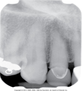
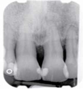
One-surface and multi-surface amalgam restorations
One-surface restorations appear as distinct, small, round or void radiopacities. They may be seen on buccal, lingual, or occlusal surfaces.
Larger two-surface and multisurface amalgam restorations also appear radiopaque and are characterized by irregular outlines or borders.
 |
 |
| One-surface restoration | Multi-surface amalgam restorations |
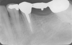
Amalgam Overhangs
Extensions of amalgam seen beyond the crown portion of a tooth in the interproximal region. Disrupts natural cleansing contours of the tooth, traps food and plaque, and contributes to bone loss.

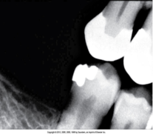
Amalgam Fragments
Fragments of amalgam may be inadvertently embedded in adjacent soft tissue during restoration of a tooth. They appear as dense radiopacities with irregular borders.
 |
 |
| Amalgam fragment seen in soft tissue near the maxillary canine | Amalgam tattoo seen by the bluish pigmentation of the gingiva |
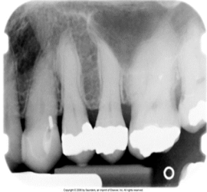
Gold Restorations
Gold restorations appear completely radiopaque and, unlike amalgam restorations, exhibit a smooth marginal outline. Gold crowns and bridges appear as large radiopaque restorations with smooth contours and regular borders. Gold foil restorations appear as small round radiopacities.
 |
 |
| Two gold crowns seen on mandibular molars. Note the smooth outline and contour. | Gold onlays seen on maxillary premolars. Note the distinct outline and contours. |
Stainless Steel and Chrome Crowns
Stainless steel and chrome crowns appear radiopaque, but not as densely radiopaque as amalgam or gold. Outlines and margins appear smooth and regular, while some areas may appear “see-through” on a radiograph.
 |
 |
 |
| Stainless steel crown seen on the mandibular first molar | Stainless steel crown area appears “see-through.” | Stainless steel crowns visible on primary teeth on this panoramic image |
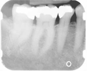
Post and Core Restorations
Post and core restorations can be seen in endodontically treated teeth, and appears radiopaque on a dental image. The core portion resembles the prepared portion of a tooth crown, and the post portion extends into the pulp canal.
Porcelain Restorations
The appearance of porcelain restorations is slightly radiopaque and resembles the radiopacity of dentin. All-porcelain crowns are a thin radiopaque line outlining the prepared tooth that represents cement may be evident through the slightly radiopaque porcelain crown. With porcelain-fused-to-metal crown, the metal component appears completely radiopaque, and the porcelain component appear slightly radiopaque.
 |
 |
| All Porcelain crown seen on the maxillary lateral incisor. Note the outline of the tooth preparation covered with radiopaque cement. | Porcelain-fused-to-metal crown seen on the maxillary central incisor. |
Composite Restorations
Composite restorations may vary in appearance from radiolucent to slightly radiopaque depending on the composition of the composite material.
 |
 |
| Composite restorations seen on maxillary anterior teeth | Composite restorations seen on mandibular anterior teeth. Note the radiolucent and radiopaque appearance of these restorations. |
Acrylic Restorations
Acrylic restorations are often used as an interim or temporary crown or filling. Acrylic is the least dense of all nonmetallic restorations and appears radiolucent or barely visible on a dental image.

Identification of Materials Used in Dentistry
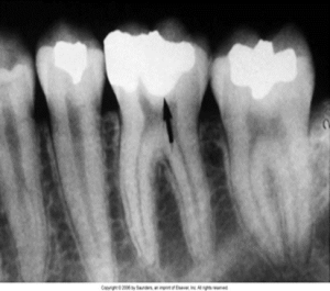
Materials used in restorative dentistry include base materials and metallic pins. Base materials are used as cavity liners placed on the floor of a cavity preparation to protect the pulp. They appear radiopaque, less radiodense than amalgam.
Metallic pins are used to enhance retention of amalgam or composite. They appear as cylindrical or screw-shaped radiopacities.
 |
 |
| Base material seen under an amalgam restoration on a mandibular first molar. | Metallic pin enhancing the retention capability of the composite restoration seen in this maxillary canine. |
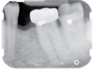
Materials used in endodontics include gutta percha and silver points. Gutta percha is a claylike material used to fill pulp canals, and appears radiopaque, similar to base materials, is less radiodense than metallic restorations.
Silver points are used to fill pulp canals, and are very radiopaque, similar to other metallic materials, appear more radiodense than gutta percha.
 |
 |
| Gutta percha seen in the pulp canal of a mandibular first molar. | Silver point seen in the pulp canal underneath a porcelain-fused-to-metal crown on the mandibular first molar. |
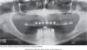 |
 |
| Complete Maxillary Denture | Complete Maxillary & Mandibular Dentures |
Materials used in orthodontics include orthodontic bands, brackets, and wires, and may be observed on dental images. They have a characteristic appearance.
 |
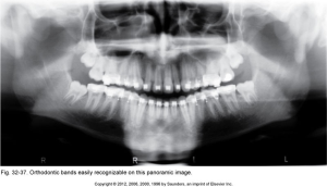 |
| Orthodontic Appliance | Orthodontic Brackets and Wires |
Implants are being used with increased frequency in oral surgery. The appearance varies based on the shape and design of the implant used.
Suture wires, metallic splints and plates, bone screws, and stabilizing arches are used in oral surgery to stabilize fractures of the maxilla and mandible.
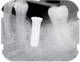 |
 |
| Implant placed in the area of the mandibular left first molar. | An implant is placed in the area of the mandibular left first molar. |
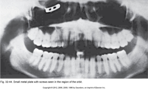 |
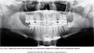 |
| Small metal plate with screws seen in the region of the orbit. | Stabilizing arches and wires seen on a panoramic image from a patient with a mandibular fracture. |
Identification of Miscellaneous Objects
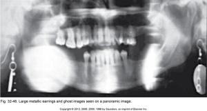
Jewelry such as metal earrings appear as dense radiopacities. A radiodense object causes an artifact known as a ghost image. These images can obscure important information and render the image nondiagnostic.
Necklaces and nose jewelry may also be seen.
 |
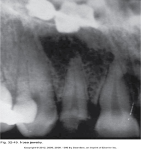 |
| Large metallic earrings and ghost images seen on a panoramic image | Nose jewelry |
The metal portion of eyeglass frames appears as a radiopacity on intraoral and extraoral dental images.

Napkin chains may be seen on an extraoral image.
If they contain any metal components, hearing aids should be removed prior to exposure of extraoral images
Shrapnel or small metal fragments that scatter outward from an exploding device may be viewed on dental images.
