31.1
Why Use Descriptive Terminology?
Descriptive terminology allows dental professionals to describe and discuss what is seen on a dental image intelligently and to communicate using a common language. It eliminates the chance for miscommunication and allows for documentation that images were reviewed.
Descriptive terminology is terms used to describe the appearance, location, and size of a lesion. This information should be documented for all lesions viewed on dental images.
Interpretation defined in Chapter 30, is the ability to read what is revealed by a dental image. To interpret dental images, the dental professional must be able to describe what is observed in accurate and succinct terms.
Descriptive Terminology versus Diagnosis
Descriptive terminology allows the dental professional to describe what is seen on an image without implying a diagnosis. It is extremely difficult, if not impossible to make a diagnosis from a dental image alone. The patient’s medical and dental history, clinical findings, signs and symptoms, laboratory tests, and biopsy results are necessary for the dentist to make a definitive diagnosis.
Radiolucent versus Radiopaque
Radiolucent |
Radiopaque |
| The portion of a processed image that is dark or black. Dental caries appears radiolucent on a dental image because the area of tooth with caries is less dense than surrounding structures. | The portion of a processed image that appears light or white. A metallic restoration appears radiopaque because it is very dense and absorbs the radiation. Two gold crowns on the mandibular molars appear radiopaque on a dental image. |
 |
 |
Radiolucent Lesions
Unilocular radiolucent lesions have one compartment and tend to be small and nonexpansile (not capable of expansion). They have borders that may appear corticated or noncorticated on image.
Unilocular radiolucent lesion with corticated borders |
Unilocular radiolucent lesion with noncorticated borders |
| The lesion exhibits a thin, well-demarcated radiopaque rim of bone at the periphery, and is usually indicative of a benign, slow-growing process. | The lesion does not exhibit a thin radiopaque rim of bone at the periphery. The periphery appears fuzzy or poorly defined, and may represent either a benign or a malignant process. |
 |
 |
Multilocular radiolucent lesions exhibits multiple radiolucent compartments. They generally have well-defined, corticated margins and are frequently expansile (capable of expansion). These lesions are typically benign with aggressive growth potential.
Multilocular radiolucent lesions may appear in a periapical location, inter-radicular location (between the roots of adjacent teeth), edentulous location or a pericoronal location (around the crown of a tooth). They may also appear as alveolar bone loss.
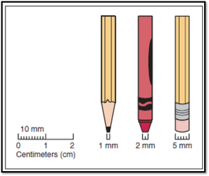
Radiolucent lesions can vary in size from several millimeters in diameter to several centimeters in diameter. They can be measured on an image with a millimeter ruler. Documentation of size is important for treatment as well as for future comparisons.
Radiopaque Lesions
Radiopaque lesions can be described as one of the following terms:
-
- focal opacity
- target lesion
- multifocal confluent
- Irregular
- ground glass
- mixed lucent-opaque
Radiopaque lesions occur not only in bone but in soft tissue as well.
-
- “Soft-tissue radiopacity”
