Normal Anatomic Landmarks
Normal anatomic landmarks of the mandible include the bony landmarks of maxilla and surrounding structures, bony landmarks of mandible and surrounding structures, air spaces seen on panoramic images, and soft tissues seen on panoramic images.
Bony Landmarks of the Maxilla and Surrounding Structures
- Mastoid process
- Styloid process
- External auditory meatus
- Glenoid fossa
- Articular eminence
- Lateral pterygoid plate
- Pterygomaxillary fissure
|
- Maxillary tuberosity
- Infraorbital foramen
- Orbit
- Incisive canal
- Incisive foramen
- Anterior nasal spine
- Nasal cavity
|
- Nasal septum
- Hard palate
- Maxillary sinus and floor of maxillary sinus
- Zygomatic process of maxilla
- Zygoma
- Hamulus
|
Radiopaque or Radiolucent Maxillary Landmarks
- Radiopaque resists the passage of the x-ray beam and limits the amount of x-rays that reach the receptor and will appear light or white.
- Radiolucent structure readily permits the passage of the x-ray beam and allows more x-ray to reach the receptor and will appear dark or black.
 |
| 1. External auditory meatus 2. Pterygomaxillary fissure 3. Infraorbital foramen. 4. Orbit 5. Anterior nasal spine 6. Nasal septum 7. Nasal conchae 8. Hard palate 9. Zygomatic process of maxilla |
 |
| 1. Glenoid fossa 2. Articular eminence 3. Maxillary tuberosity 4. Maxillary sinus zygoma |
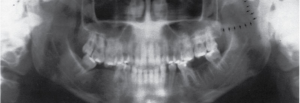 |
| Lateral pterygoid plate |
Bony Landmarks of Mandible and Surrounding Structures
- Mandibular condyle
- Coronoid notch
- Coronoid process
- Mandibular foramen
- Lingula
- Mandibular canal
|
- Mental foramen
- Hyoid bone
- Mental ridge
- Mental fossa
- Lingual foramen
- Genial tubercles
|
- Inferior border of the mandible
- Mylohyoid ridge
- Internal oblique ridge
- External oblique ridge
- Angle of the mandible
|
 |
|
1. condyle 2. coronoid notch 3. coronoid process 4. mandibular foramen 5. mental foramen 6. genial tubercles 7. styloid process
|
Air Spaces Seen on Panoramic Images
 |
| Air spaces seen on panoramic images: 1. palatoglossal air space 2. nasopharyngeal air space 3. glossopharyngeal air space |
Soft Tissues Seen on Panoramic Images
 |
|
Soft tissues seen on panoramic images: 1. tongue 2. soft palate and uvula 3. lipline 4. ear
|
Media References:
- Images courtesy of Iannucci and Howerton: Dental Radiography Principles and Techniques, 6th Edition, (2022), Chapter 29.

