28.3
Bony Landmarks of the Mandible
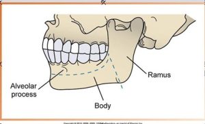
The largest and strongest bone of the face, the mandible is the only moveable bone of the face. The mandible is divided into three main parts:
- Ramus
- Vertical portion found posterior to the third molar
- Angle of the mandible is at the corner portion of the ramus
- Body of the Mandible
- Horizontal U-shaped portion from ramus to ramus
- Alveolar process
- Encases and supports the teeth
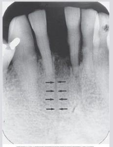
Genial Tubercles
Tiny bumps of bone on the lingual aspect of the mandible. Attachment sites for genioglossus and geniohyoid muscles. Genial tubercles appears as a ring-shaped radiopacity below the apices of the mandibular incisors
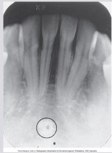
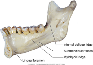
Lingual Foramen
A tiny opening or hole in bone located on the internal surface of the mandible. The lingual foramen appears as a small radiolucent dot inferior to the apices of the mandibular incisors. The lingual foramen is located near the midline and is surrounded by the genial tubercles, which appear as a radiopaque ring.
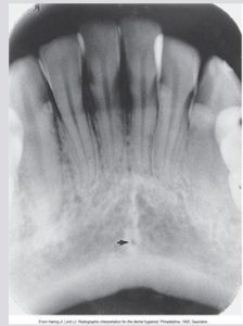
Nutrient Canals
Tubelike passageways through bone containing nerves and blood vessels that supply the teeth. Most often seen in anterior mandible. Nutrient canals appear as thin, vertical radiolucent lines readily seen in areas of thin bone.
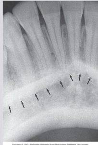
Mental Ridge
A linear prominence of cortical bone located on the external surface of the anterior portion of the mandible. The mental ridge extends from the premolar region to the midline and slopes slightly upward. The mental ridge appears as a thick radiopaque band that extends from the premolar region to the incisor region. Often appears superimposed over the mandibular anterior teeth.
 |
 |
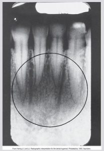
Mental Fossa
A scooped-out, depressed area of bone located on the external surface of the anterior mandible. The mental fossa is located above the mental ridge in the mandibular incisor region. A ________________ area above the mental ridge. The radiographic appearance of the mental fossa varies and is determined by the thickness of the bone in the anterior region of the mandible.
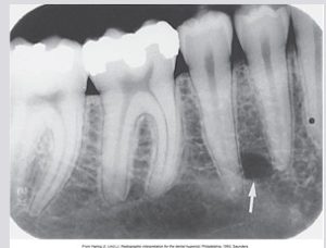
Mental Foramen
An opening or hole in bone located on the external surface of the mandible in the region of the mandibular premolars. Blood vessels and nerves that supply the lower lip exit through the mental foramen. The mental foramen appears as a small ovoid or round radiolucent area located in the apical region of the mandibular premolars. Frequently misdiagnosed as a periapical cyst, granuloma, or abscess because of its location.
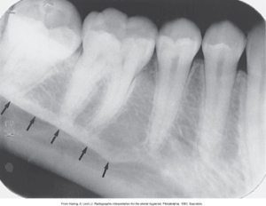
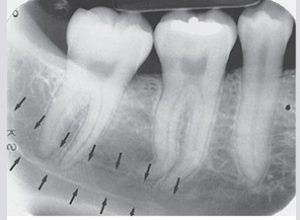
Mylohyoid Ridge
A linear prominence of bone located on the internal surface of the mandible. Extends from the molar region downward and forward toward the lower border of the mandibular symphysis. The mylohyoid ridge appears as a dense radiopaque band that extends downward and forward from the molar region. May appear to be continuous with the internal oblique ridge.
 |
 |
Mandibular Canal
A tubelike passageway through bone that travels the length of the mandible. Houses the inferior alveolar nerve and blood vessels. Extends from the mandibular foramen to the mental foramen. The mandibular canal appears as a radiolucent band outlined by two thin radiopaque lines that represent the cortical walls of the canal.
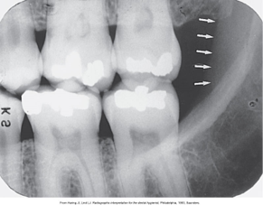
Anterior Border of the Ramus
Anterior border of the ramus extends vertically downward from the coronoid process to the external oblique ridge. Also known as the internal oblique line. On a molar bite-wing image, the descending ramus of the mandible may be seen as a slightly radiopaque vertical band posterior to the maxillary and mandibular molars.
 |
|
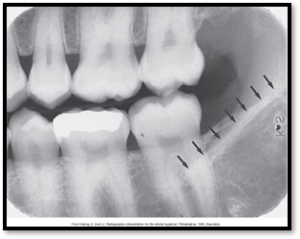
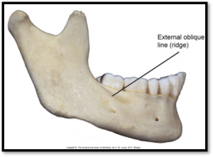
External Oblique Ridge
Also known as the external oblique line, the external oblique ridge is a linear prominence of bone located on the external surface of the body of the mandible. The external oblique ridge appears as a radiopaque band extending downward and forward from the anterior border of the ramus of the mandible. Typically ends in the mandibular 3rd molar region. The anterior border of the ramus ends in the external oblique ridge.
 |
 |
Submandibular Fossa
Also known as mandibular fossa or submaxillary fossa, the submandibular fossa is a scooped-out, depressed area of bone located on the internal surface of the mandible inferior to the mylohyoid ridge.The submandibular fossa appears as a radiolucent area in the molar region below the mylohyoid ridge. May be slightly more radiolucent than the adjancent bone on some periapical radiographs. The submandibular salivary gland is located in the submandibular fossa.
 |
|
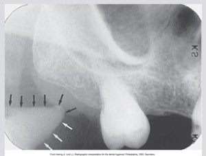
Coronoid Process
A marked prominence of bone on the anterior ramus of the mandible with a radiographic appearance. The coronoid process appears as a triangular radiopacity superimposed over, or inferior to, the maxillary tuberosity region. Not seen on a mandibular periapical but does appear on a maxillary molar periapical. It serves as an attachment site for one of the muscles of mastication.