27.2
Step-by-Step Procedures
It is critical to refer to the manufacturer-provided instruction booklet for information concerning the system’s operation, equipment preparation, patient preparation, and exposure.
Patient Preparation
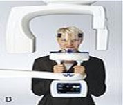
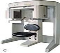
Patients may be asked to sit or stand or be placed in the supine position during radiation exposure depending on manufacturer instructions as the number and size of the machine varies with different manufactures. Instructions are given to the patient before exposure to remove jewelry, eyeglasses, and removable dental appliances. A guide may be placed in the patient’s mouth during the scanning process and some specialists may ask that the upper and lower teeth be kept apart.
Patient Positioning
For patient positioning, the patient should be instructed to remain still as scan times can vary from 7 to 30 seconds and the ergonomic head and chin supports have been designed for improved patient comfort. Laser beams may be installed to help with the proper alignment of clinical structures and to ensure correct anatomic positioning.
 |
 |
 |
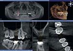
| A CBCT standing machine. | A CBCT sitting machine. | Diagnostic capabilities of three-dimensional cone beam imaging showing implant placement of tooth 1.6 |
Advantages & Disadvantages of Three-Dimensional Digital Imaging
The advantages of three-dimensional digital imaging is:
- Lower radiation dose: The amount of radiation from a typical CBCT scan compared with the dose given for three or four full-mouth series of intraoral radiographs
- Brief scanning time: The quick 8-10 second scan decreases chance of motion artifacts and increases high level of patient compliance
- Anatomically accurate images: Provides an accurate measurement of anatomic structures with a 1.1 ratio relationship
- Ability to save and easily transport images: Three-dimensional images can be saved and viewed online, e-mailed, printed, or placed on a compact disc.
- The amount of radiation from a typical CBCT scan compared with the dose given for three or four full-mouth series of intraoral radiographs.
- Three-dimensional images can be saved and viewed online, e-mailed, printed, or placed on a compact disc.
The disadvantages of three-dimensional digital imaging is:
- Patient movement and artifacts
- Size of the field of view
- Cost of equipment: CBCT machines currently range in cost from $80,000 to $175,000
- Lack of training in the interpretation of images on data on areas outside the maxilla and the mandible
- Training must be completed to use equipment properly
- Only trained dentist registered with the Royal College of Surgeons of Ontario and with specialized training in CBCT can place, expose and diagnose cone beam computed technology within Ontario scans
-

