20.3
Step-by-Step Procedures
The dental radiographer should have the radiography operatory and equipment prepared before seating the patient. A definite order should be used when taking radiographs to prevent errors, and time should be used efficiently. The following steps are:
- Patient Preparation
- Equipment Preparation
- Exposure Sequence for Receptor Placements
- Receptor Placement for Bisecting Technique
Patient Preparation
Patient preparation includes infection control procedures, preparation of treatment area and supplies, and the patient is seated and prepared. The patient should be seated upright and both arches of the mouth should be parallel to the floor.
The you will move on to explain the radiographic procedure to the patient and position them in the chair. You will likely need to adjust the headrest and the place secure the lead apron with a thyroid collar and remove all objects from the patient’s mouth. The midsaggital plane should be perpendicular to the floor and the patient’s eyeglasses must also be removed.
Equipment Preparation
For preparing the equipment you will need to set the exposure factors and open the sterilized package containing the beam alignment device if beam alignment devices are used.
Exposure Sequence for Receptor Placements
The anterior exposure sequence begins with the maxillary right canine tooth. All of the maxillary anterior teeth will be exposed and end with the maxillary left canine. Then move to the mandibular arch by beginning with the mandibular left canine. Expose all of the mandibular anterior teeth and finish with the mandibular right canine.
When taking radiographs using the bisecting technique, always begin with the anterior teeth. Working from left to right in the mandibular arch, no wasted movement or shifting of the PID occurs.
The hotspot below highlights the prescribed placements for anterior periapicals.
The posterior exposure sequence begins with the maxillary right quadrant. Then move to the mandibular right quadrant and move to the maxillary left quadrant. Finish with the mandibular left quadrant. In each quadrant, expose the premolar receptor first and then the molar receptor.
The hotspot below highlights the prescribed placements for posterior periapicals.
Receptor Placement for Bisecting Technique
Receptor placement is the specific area where the receptor must be positioned before exposure and is dictated by the teeth and surrounding structures that must be included on the resultant radiograph.
| Anterior placement includes: | Posterior placement includes: |
|
|
Guidelines for Receptor Placement and Bisecting Technique
Posterior receptors are always placed horizontally and anterior receptors are always placed vertically. The white side of the receptor always faces the teeth. The incisal or occlusal edge of the receptor must extend approximately 1/8 inches beyond the teeth. Remember to always center the receptor over the area to be examined.
Advantages and Disadvantages
The advantages of receptor placement and bisecting technique are it can be used without a beam alignment device (shallow palate, bony growth, sensitivity) and decreases exposure time with the use of a short PID (8 inches). The two disadvantages are image distortion and angulation problems.
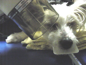
Below is an image of an example of a bisecting angle technique for the upper premolars being done on an adorable furry friend.
Practice Makes Perfect
Media Attributions
- Iannuci: Dental Radiography, 6th Edition, Chapter 20, CC BY-NC-ND
