2.2
X-Radiation & X-Rays
X-radiation is high-energy, ionizing electromagnetic radiation. X-rays are weightless bundles of energy without an electrical charge that travel in waves with a specific frequency at the speed of light. X-ray photons interact with the materials they penetrate and cause ionization.
Properties of X-rays
Property |
What it is |
| Appearance | Are Invisible and cannot be directed by any of the senses |
| Mass | No weight |
| Charge | No charge |
| Speed | Travel at the speed of light |
| Wavelength | Travel in waves and have short wavelengths with high frequency |
| Path of travel | Travel in straight lines and can be deflected or scattered |
| Penetrating power | Can penetrate liquids, solids, and gases. The composition of the substance determines whether x-rays penetrate or pass through, or are absorbed. |
| Absorption | Are absorbed by matter; the absorption depends on the atomic structure of matter and the wavelength if the x-ray |
| Ionization capabilities | Interact with materials they penetrate and cause ionization |
| Fluorescence capability | Can cause certain substances to fluoresce or emit radiation in longer wavelengths (e.g.: visible light and ultraviolet light) |
| Effect on receptor | Can produce an image on a receptor |
| Effect on living tissues | Can cause biologic changes in living cells |
| Focusing capability | Cannot be focused to a point and always diverge from a point |
Component Parts
The component parts of an x-ray machine is control panel, extension arm, and tubehead.
Control Panel
The control panel contains an on-off switch, an indicator light, and control devices: time, kilovoltage, and milliamperage. It is plugged into an electrical outlet.
Extension Arm
The extension arm component suspends the x-ray tubehead, houses the electrical wires that extend from the control panel to the tubehead and allows for movement and positioning of the tubehead.
Tubehead
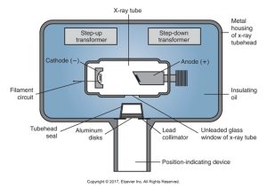
A tubehead is a tightly sealed, heavy metal housing that contains an x-ray tube that produces dental x-rays.
The metal housing surrounds the x-ray tube and transformers protects the tube, and grounds high-voltage components. The insulating oil surrounds x-ray tube and transformers, preventing overheating.
The tubehead seal permits exit of x-rays from tubehead, seals the oil, and filters x-ray beam where the x-ray tube is the heart of the generating system. And the transformer alters voltage of incoming electricity.
The following images show more important components of a Tubehead.
Below is a diagram of an x-ray tubehead and its internal components, including the x-ray tube, cathode, anode, transformers, and lead collimator, all enclosed in a metal housing with insulating oil.
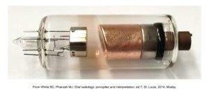
X-Ray Tube
A glass vacuum tube measures several inches long by 1 inch in diameter and includes leaded-glass housing, cathode, and anode.
This is an image of an x-ray tube component from dental radiographic equipment with visible metal and glass parts.
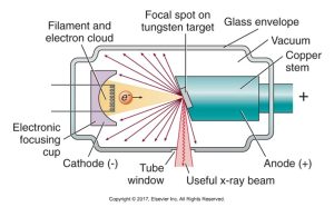
Leaded-Glass Housing
A leaded-glass vacuum tube prevents x-rays from escaping in all directions. One area has a “window” that permits the x-ray beam to exit the tube and directs the beam toward the aluminum disks, lead collimator, and PID.
Cathode (−)
A cathode is a negative electrode that consists of a tungsten wire filament in a cup-shaped holder made of molybdenum. The cathode supplies the electrons necessary to generate x-rays. The tungsten filament produces electrons when heated, and the molybdenum cup focuses electrons into a narrow beam and directs the beams toward the tungsten target.
Anode (+)
An anode is a positive electrode that consists of a wafer-thin tungsten plate embedded in a solid copper rod. The anode converts electrons into x-ray photons. The tungsten target serves as a focal spot and converts electrons into photons, and the copper stem functions to dissipate heat away from the tungsten target.
The production of dental x-rays occurs in the x-ray tube.
The diagram below shows the internal structure of an x-ray tube, including the filament and electron cloud, electronic focusing cup, cathode, anode, focal spot on the tungsten target, glass envelope, and the direction of the useful x-ray beam through the tube window.
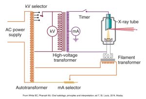
X-Ray Generating Apparatus
An x-ray generating apparatus is in 3 sections: Electricity and electrical currents, circuits, and transformers.
The three different transformers that are used in the production of dental x-rays: step-down transformer, step-up transformer, and autotransformer.
The diagram below displays an x-ray machine circuit, showing the AC power supply, autotransformer, kV and mA selectors, high-voltage transformer, timer, filament transformer, and the X-ray tube.
Practice Makes Perfect
Drag and drop the component name to its proper place on the x-ray tubehead diagram.
Drag and drop the component name to its proper place on the x-ray tubehead diagram.
Media Attributions
- Iannucci & Howerton: Dental Radiography Principles and Techniques, 6th Edition, Chapter 2, CC BY-NC-ND
