18.2
Diagnostic Criteria for Intraoral Images
Images must have optimum density, contrast, definition, and detail and also must have the least amount of distortion possible. The CMS must include images that show all tooth-bearing areas and periapical images must show the entire crowns and roots of teeth being examined, as well as 2 to 3 mm beyond the root apices. Bite-wing images must show open contacts.
In the Iannucci & Howerton, Dental Radiography Principles & Techniques, 6th Edition textbook, on page 165, refer to Box 18-1 General Diagnostic Criteria for intraoral Images, and Pg. 164 Fig 18-4: complete mouth series.
Extraoral Imaging Examination
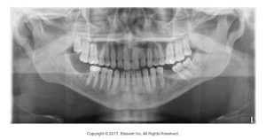
An extraoral imaging examination is an inspection of large areas of the skull or jaws. Extraoral receptors are receptors that are placed outside the mouth. Some examples of common extraoral images is a panoramic image, lateral jaw, lateral cephalometric, posteroanterior, and waters.
In the Iannucci & Howerton, Dental Radiography Principles & Techniques, 6th Edition textbook, on page 165, refer to figure 18-5 and the helpful hint box: extraoral imaging exam.

Prescribing Dental Images
Prescribing is based on the individual needs of the patient, and the dentist uses professional judgment to make decisions about the number, type, and frequency of dental images. All images are prescribed based on the patient’s needs and not their insurance coverage.
In the Iannucci & Howerton, Dental Radiography Principles & Techniques, 6th Edition textbook, on pages 34-35, refer to table 4-1 Recommendations for Prescribing Dental Radiographs (2012) for guidelines.
Not all patients need a CMS. CMS is appropriate when a new adult patient presents with clinical evidence of generalized dental disease or a history of extensive dental treatment; otherwise, a combination of bitewings, selected periapical, and/or a panoramic image should be prescribed on the basis of a patient’s individual needs.
Media Attributions
- Iannuci: Dental Radiography, 6th Edition, Chapter 18, CC BY-NC-ND

