18.1
Intraoral examinations are the foundation of dental imaging, which is the inspection of teeth and intraoral adjacent structures and requires the use of intraoral image receptors. An intraoral receptor is a receptor that is placed inside the mouth and is used to examine the teeth and supporting structures. The intraoral film is the most commonly used X-ray film.
Types of Intraoral Imaging Examinations
The different types of intraoral imagining examinations are a periapical examination, an interproximal examination, and an occlusal examination. Each of these examinations has a particular purpose and requires the use of a specific type of imaging receptor and technique.
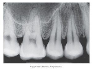
Periapical Examination
The purpose of a periapical examination is to examine the entire tooth from crown to root and supporting bone. The type of imaging receptor used is a periapical receptor. The word part peri means “around,” and the word part apex refers to the end of the tooth root. The technique is paralleling and bisecting. Below is an example of a periapical examination x-ray.
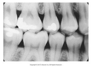
Interproximal Examination
An interproximal examination examines the crowns of the maxillary and mandibular teeth on a single film. It is often taken to check for cavities between teeth and show the crestal bone. The type of imaging receptor is the bite-wing receptor. The technique used is the bite-wing technique. Below is an image of an interproximal examination x-ray.
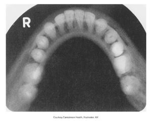
Occlusal Examination
The purpose of an occlusal examination is to examine large areas of the maxilla or the mandible on one film. The type of imaging receptor used is an occlusal receptor. This type of film is most commonly seen in pediatric dental offices. The occlusal technique is used. Below is an example of an occlusal examination x-ray.
Complete Mouth Series/Full Mouth Series
CMS, also known as FMS or FMX, is a full-mouth series or complete series that detects tooth-bearing, dentulous, and edentulous areas. Below is an example of a full mouth series of dental x-rays displaying various angles of all teeth.
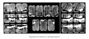
CMS/FMS/FMX series can include only periapicals or be a combination of periapicals and bite-wings. A total of 14 to 20 films may be taken, and the film size selection is important. It is used to detect disease, foreign objects, and retained roots, and patients usually have a full-mouth series of x-rays taken every 3 to 5 years.
Media Attributions
- Iannuci: Dental Radiography, 6th Edition, Chapter 18, CC BY-NC-ND
