11.1
Basic Concepts
Mount: is to place in an appropriate setting, as for display or study.
Dental radiography mounting: the placement of radiographs in a supporting structure or holder.
Mounting Images
Who mounts images? The answer to this question is that any trained dental professional (i.e., dentist, dental hygienist, dental assistant) who possesses knowledge of the typical anatomic landmarks of the maxilla, mandible, and related structures is qualified to mount images. Usually, mounting images is the responsibility of the office dental radiographer.
Images should be mounted immediately after processing and should be done in an area designated for mounting.
Using a mount is quicker and easier to view and interpret, is easily stored and available for interpretation, and decreases the chances of error in determining the patient’s right and left side. Most digital imaging systems allow the dental radiographer to choose the appropriate-size mount, and the mounts should be labeled with the patient’s full name and date of exposure.
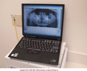
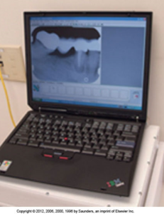
Below are images of digital images of the mandibular right periapical region in a mount and panoramic images as soon as possible on a computer monitor.
 |
 |
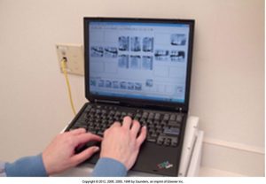
See the following image of a full mouth series of radiographs exposed using digital imaging. Each image is placed in the appropriate window on the mount and arranged in anatomic order.

Media Attributions
- Iannucci & Howerton Principles and Techniques: Dental Radiography, 6th Edition, Chapter 11, CC BY-NC-ND

