1.2

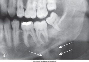
In dentistry, dental images enable the dental professional to identify many conditions that may otherwise go undetected and to see conditions that cannot be identified clinically.

A dental X-ray showing the side view of a jaw with arrows pointing to specific teeth, possibly indicating areas of interest or concern.

Uses of Dental Imaging

Dental images are utilized to detect lesions, confirm or classify suspected diseases, and localize lesions or foreign objects. They provide valuable information during dental procedures, evaluate growth and development, and illustrate changes secondary to caries, periodontal disease, and trauma. Furthermore, dental images document the condition of a patient and aid in the development of a clinical treatment plan.

Roentgen and the Discovery of X-Rays

Wilhelm Conrad Roentgen – 1895

Black and white portrait of Wilhelm Conrad Röntgen, the discoverer of X-rays, with a distinguished beard and mustache, alongside an early X-ray tube apparatus.

Roentgen was experimenting with vacuum tubes and fluorescent screens. He noticed the screens were glowing even though they were several feet from the tube, and he determined that some mysterious ray was causing them to glow.

The image below is the first radiograph of the human body showing Roentgen’s wife’s hand.

The first radiograph of Roentgen’s wife's hand

Earlier Experimentation & Pioneers in Dental X-Radiation Timelines

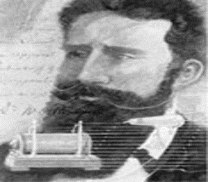
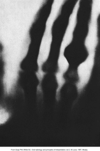
Not mentioned in the timeline for pioneers in dental x-radiation, Willaim H. Rollins (n.d.) developed the first dental x-ray unit. Frank Van Woert (n.d.) was the first to use film in intraoral radiography.

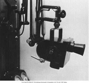
History of Dental X-Ray Equipment

Not mentioned in the timeline of dental x-ray equipment, Victor CDX shockproof tube housing was developed in 1923.

This is an image of an early 20th-century X-ray tube housing, part of medical radiographic equipment from 1923.

A black and white image of an early 20th-century X-ray tube housing, part of medical radiographic equipment from 1923.

History of Dental Film

The history of dental film spans over the years 1896 to 1913, where hand-wrapped glass photographic plates or film were developed. And in 1913, Eastman Kodak manufactured prewrapped intraoral film.

This is an image of prewrapped intraoral dental X-ray films.

A set of five prewrapped intraoral dental X-ray films in white and teal packaging, arranged diagonally.


History of Dental Radiographic Techniques

Along with the timeline, Yrjo Paatero (n.d.) experimented with a slit bean of radiography, intensifying screens, and rotational techniques.

Also not mentioned in the timeline of dental radiographic techniques, radiographs have been produced using radiographic film for well over a century. Traditional radiography is being replaced by digital imaging. Digital imaging allows for instant and easy transmission of images and electronic storage. In 1987 the first intraoral imaging sensor was developed and later in 1989 an article describing direct digital imaging technology was published in U.S. dental literature.


Media Attributions

  • Iannuci: Dental Radiography, 6th Edition, Chapter 1, CC BY-NC-ND

License

Icon for the Creative Commons Attribution-NonCommercial-NoDerivatives 4.0 International License

DE 115: Dental Radiography Copyright © 2024 by verabodnarchuk; monicacoggin; alisonloach; and rbhatia2 is licensed under a Creative Commons Attribution-NonCommercial-NoDerivatives 4.0 International License, except where otherwise noted.

Share This Book