12.1
A dental image is a photographic image that is produced on film by the passage of x-rays through teeth and supporting structures that are two-dimensional representations of a three-dimensional object. Dental images are a necessary component of comprehensive patient care.
Importance of Dental Images
Dental images are a necessary component of comprehensive care and are essential for diagnostic purposes. Images enable the dental professional to identify many conditions that may otherwise go undetected and gain a great deal of information about teeth and supporting bone structures. An oral exam limits the practitioner to a knowledge of what is only seen clinically.
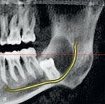
In the Iannucci & Howerton, Dental Radiography Principles & Techniques, 6th Edition textbook on page 124, refer to Figure 12-1.
Below is an image of a young adult patient presented to the dental office for a routine preventive appointment. The panoramic image reveals an impacted tooth 3.7.
Dental images can be used to educate the dental patient about some of these common conditions that are only detected with the use of dental images. Information that can be found in a dental image includes missing teeth, extra teeth, impacted teeth, dental caries, periodontal disease, tooth abnormalities, retained roots, as well as cysts and tumors
Use of Dental Images
The uses of dental images includes:
- Detection of diseases, lesions, and conditions of the teeth and bones that cannot be identified clinically
- Confirming suspected diseases
- Assisting in the localization of lesions and foreign objects
- Essential component of the patient record
- Follow-up images can be compared with initial images and examined for changes
Benefits of Dental Images
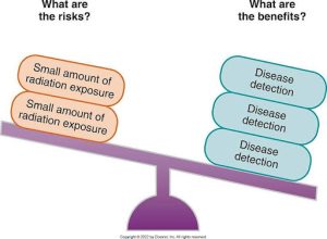
The primary benefit of dental images is the detection of disease. This benefit far outweighs the risk of small doses of x-radiation, and its use assists dental professionals in identifying and preventing problems, such as tooth-related pain or the need for surgical procedures. Dental images can minimize and prevent future problems. There are potential risks that can be involved, but the benefits usually outweigh those odds.
Below is a graphic displaying the comparison of the risks and benefits of dental images. The risks are small amounts of radiation exposure, and the benefits are disease detection.
Media Attributions
- Iannucci & Howerton: Dental Radiography Principles and Techniques, 6th Edition, Chapter 12, CC BY-NC-ND
