11.2
Image Mounting Methods
Identification of landmarks helps distinguish maxillary periapical images from mandibular periapical images. It is essential to note the curve of spee when mounting bite-wing radiographs.
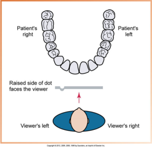
An identification dot is located in one corner of each intraoral film packet that is a small and raised bump. All the dots must face the same direction when mounted and the film is positioned in the packet such that the raised side of the dot faces the x-ray beam during exposure. It is used to determine film orientation.
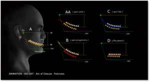
Below is an image of a head profile displaying the jaw and the curve of spee, with four different occlusal plane adjustments depicted by colored lines and teeth, illustrating variations in the curvature from early to late apex positions.
Labial mounting is when radiographs are placed in the film mount with the raised (convex) side of the identification dot facing the viewer (dental radiographer). Lingual mounting is radiographs that are placed in the film mount with the depressed (concave) side of the identification dot facing the viewer (dental radiographer).
 |
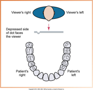 |
| With the labial mounting method, the radiographs are viewed as if the dental radiographer were looking directly at the patient. | With the lingual mounting method, the radiographs are inside the patient’s mouth a viewed as if the dental radiographer nd looking out. |
Step-By-Step Procedure
The suggested mounting order is:
- Bite-wings
- Maxillary anterior periapicals
- Mandibular anterior periapicals
- Maxillary posterior periapicals
- Mandibular posterior periapicals
Then check the radiographs by ensuring all embossed dots (pimple not dimple) are oriented correctly, all the images are properly arranged in anatomical order, and that the mount is properly dated and saved. To help identify maxillary from mandibular teeth your knowledge of dental anatomy is important. Remember maxillary teeth have 3 roots and the root tips appear to flare towards the left or right depending on the the side of the exposure. The mandibular molars generally have 2 very distinct 2 roots. Images are mounted in the mount with maxillary roots facing the ceiling and mandibular roots facing the floor.
Bite-Wing, Anterior & Prosterior Periapicals
 |
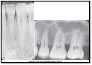 |
| A bite-wing image shows the crowns of both maxillary and mandibular teeth on one film. | A. An anterior periapical film is oriented vertically.
B. A posterior periapical film is oriented horizontally. |
Maxillary and Mandibular Image Mounts
Order of Teeth
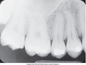
The order of teeth can be used to distinguish right from left; premolars are located in front of molars; therefore, below is a maxillary right periapical image.
Practice Makes Perfect
Media Attributions
- Iannucci & Howerton: Dental Radiography Principles & Techniques, 6th Edition, Chapter 11, CC BY-NC-ND
