6.1
A dental radiograph appears as a black-and-white image or picture with varying shades of gray.
Radiolucent is the portion of the radiograph that is dark or black and a structure that appears black on a radiograph that lacks density.
Radiopaque is the portion of the radiograph that appears white and a structure that appears white on a radiograph that is dense and absorbs or resists the passage of the x-ray beam.
In a diagnostic radiograph the images have proper density and contrast, sharp outlines, and are of the same shape and size as the object radiographed. Dental radiographs must be of diagnostic quality in order for the dentist to provide proper quality care
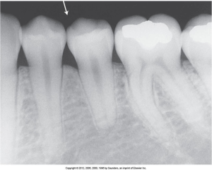
Radiolucent Air Space
The image below, where the arrow is pointing, is where air space appears radiolucent or dark because the dental x-rays pass through freely.
Enamel, dentin, and bone are dense structures. These areas resist the passage of x-rays and appear radiopaque, or white. Although dentin is more radiopaque than dental pulp, it is more radiolucent than the overlying enamel. In order to ensure that these differences are noticeable, both contrast and density must be optimized.
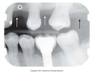
Gray Area
Note that in the image below, there is a grayish area in the upper arch where the arrows are pointed; this represents the gingival tissues.
Visual Characteristics
Density and contrast are the two visual characteristics that influence the quality of a dental radiograph. It is important to know how to alter radiographic variables in order to change the contrast, density, and image detail.
Density
Density is the overall darkness or blackness of a dental radiograph. Some influencing factors are milliamperage, kilovoltage, exposure time (if PIDs are changed on a tubehead, it is important to adjust the exposure time accordingly), and the subject thickness.
Contrast
Contrast is the difference in degrees of blackness between adjacent areas. The difference in the amount of light transmitted through adjacent areas of a dental image. A radiographic image that is a compromise between low contrast and high contrast is preferred.
Influencing factors of contrast is the increasing the kilovoltage affects image contrast by increasing the mean or average energy of the x-rays and by producing higher energy x-rays. The more areas of varying tissue density are recorded on the image and appear as shades of gray. And remember, kilovoltage controls the contrast.
kVp, Density, and Contrast
Media Attributions
- Iannucci & Howerton: Dental Radiography Principles and Techniques, 6th Edition, Chapter 6, CC BY-NC-ND
