35.2
Pulpal Lesions Viewed on Dental Images
Examination of the pulp chambers and canals is impossible without dental images. Many dental procedures require information about the size and location of the pulp cavity before treatment begins. Without dental images, examination of pulp chambers and canals is impossible.
Dental images may detect conditions such as:
-
- Pulpal sclerosis
- Pulpal obliteration
- Pulp stones
Periapical Lesions Viewed on Dental Images
A periapical lesion is located around the apex (tip of the root) of a tooth.
Periapical Radiolucencies
Periapical granulomas, cysts, and abscesses are commonly seen on dental images. These lesions cannot be diagnosed on their dental image appearance alone. Diagnosis is based on clinical features and dental image and microscopic appearance.
 |
 |
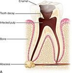
| Infection of the pulp results in necrosis. A periapical granuloma, cyst or abscess forms at the apex of the root. | Image shows infection of the pulp of tooth. |
Periapical Radiopacities
The following are a few of the common periapical radiopacities that may be seen on dental images found near tooth apdex:(?)
-
- Condensing osteitis
- Sclerotic bone
- Hypercementosis
These may be diagnosed based on their appearance, clinical information, and patient history
Media Attributions
- Images courtesy of Iannucci and Howerton, Dental Radiography Principles and Techniques,6th edition, 2022, Chapter 35.
