35.1
Trauma Viewed on Dental Images
Trauma is an injury produced by an external force. It may affect the crowns and roots of teeth as well as alveolar bone. It may also result in injuries of teeth and bone and injuries such as intrusion, extrusion, and avulsion. Trauma may result in fractures of the jaws and teeth as well as displacement of teeth.
Trauma viewed on dental images include fractures and injuries.
Fractures
Fractures are the breaking of a part and may affect the crowns and roots of teeth or the bones of the maxilla or mandible. Whenever a fracture is evident or suspected, image examination of the injured area is necessary.
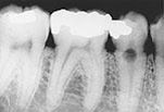
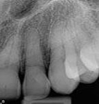
Crown Fractures most often involves anterior teeth and may involve enamel, dentin, and/or pulp. The dental image permits evaluation of the proximity of the damage to the pulp chamber and for evaluation of the root for any additional fractures
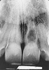
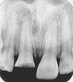
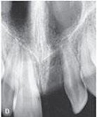
Root fractures most often occur in the maxillary central region. They are less common than crown fractures and result from an accident or traumatic blow. They may be vertical or horizontal, single or multiple
If the x-ray beam is parallel to the plane of the fracture, it will appear as a radiolucent line. If the x-ray beam is not parallel to the plane of the fracture, it may not be apparent at all
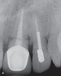
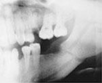
Jaw fractures are most often observed in the mandible. The panoramic image is the best film for visualizing mandibular fractures. On a dental image, the fracture appears as a radiolucent line. Maxillary fractures are typically difficult to detect on dental images.
 |
  |
 |
|
| Crown fracture | Root fracture | Jaw fracture | |
Injuries
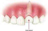
Trauma may result in the displacement of teeth. Displacement involves luxation (intrusion and extrusion) and avulsion. Dental images allow for the evaluation of structures after tooth displacement.
Luxation
Luxation is the abnormal displacement of teeth. Intrusion is the abnormal displacement of teeth into bone. Extrusion is the abnormal displacement of teeth out of bone. Teeth that have been luxated should be evaluated by a periapical image and examined for root and adjacent alveolar bone fractures, damage to the periodontal ligament, and pulpal problems.
 |
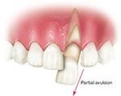 |
| An Intruded tooth | A partial evulsion |
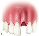
Dental Avulsion
Dental avulsion is the complete displacement of a tooth from alveolar bone. The periapical image shows a tooth socket without a tooth. Dental avulsion is the result from trauma associated with an assault or accidental fall.
Dental images are important in the evaluation of the socket areas and should be used to examine the region for splintered bone.
 |
 |
| Total avulsed tooth | (What does this figure show?) |
Resorption Viewed on Dental Images
Physiologic resorption is a process seen with the normal shedding of primary teeth. The primary tooth is shed when resorption of the roots is complete.
Pathologic resorption is a regressive alteration of tooth structure observed when a tooth is subjected to abnormal stimuli. The pathologic resorption of teeth can be described as external or internal, depending on the location of the resorption process.
External Resorption
Seen along the periphery of the root surface, external resorption is often associated with reimplanted teeth, abnormal mechanical forces, trauma, chronic inflammation, tumors and cysts, impacted teeth, or idiopathic causes. It most often involves the apices of teeth. The apical region appears blunted and the length of the root is shorter than normal. It is not detected clinically and does not have mobility, with no effective treatment
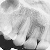
Blunted root apex
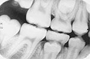
Internal Resorption
Internal resorption occurs within the crown or root of a tooth, involving the pulp chamber, pulp canals, and surrounding dentin. It is believed to be precipitated by factors such as trauma, pulp capping, and pulp polyps. It appears as a round-to-ovoid radiolucency in the midcrown or mid root portion of the tooth. Generally asymptomatic, treatment is variable.
If the tooth is weakened by the resorptive process, extraction is recommended. If the tooth has weakened, extraction may be recommended. Endodontic therapy is recommended if perforation has not occurred.