34.3
All images need to be evaluated for these two things:
- Evaluated for bone loss
- Examined for other predisposing factors that may contribute to periodontal disease
Bone Loss
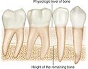
Bone loss can be estimated as the difference between the physiologic bone level and the height of remaining bone. It can be described in terms of the pattern, distribution, and severity of loss.
An image allows the dental professional to view the amount of bone remaining rather than the amount of bone lost. Bone loss estimated as the difference between the physiologic level of bone and the height of the remaining bone.
 |
 |
| Height of the remaining bone | (What does this figure show?) |
 |
 |
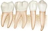
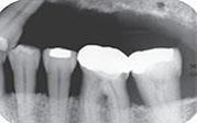
| Horizontal bone loss | Vertical bone loss |
The distribution can be described as localized or generalized. Localized bone loss occurs in isolated areas, and has less than 30% of the sites involved. Generalized bone loss occurs evenly throughout the dental arches and has more than 30% of the sites involved.
The severity of bones loss can be classified as slight, moderate, or severe. Measured by the clinical attachment loss (CAL), slight bone loss is measured at 1 to 2 mm. Moderate bone loss is measured 3 to 4 mm. Severe bone loss is 5 mm or greater. This is generally measured and charted by the dental hygienist during a clinical assessment.
The Progression of Periodontal Disease
Inflammation of the gingival tissues is typically the first sign of periodontal disease. If untreated, gingival inflammation may progress to slight or mild periodontitis. With advancing periodontal disease, horizontal or vertical bone loss may be present (distribution of the bone loss may be localized or generalized).
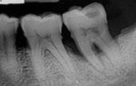
When bone in the furcation area is destroyed, a radiolucent area is evident on dental image. The bone loss associated with advanced or severe periodontitis indicates further progression of the disease and is considered severe
Predisposing Factors
Predisposing factors and local irritants may contribute to periodontal disease. Elimination is important in the management and treatment of periodontal disease. The effects of certain medications, tobacco use, and conditions such as diabetes are all considered risk factors for periodontal disease.
Dental images aid in detection of irritants such as calculus and defective restorations.
Calculus
Results from the mineralization of plaque and appears white or light on a dental image. It most often appears as a pointed or irregular radiopaque projection extending from proximal root surfaces. Stonelike concretion that forms on the crowns and roots of teeth due to the calcification of bacterial plaque.
Calculus may also appear as a:
-
- Ringlike opacity
- Nodular image projection
- Smooth opacity on a root surface
Defective Restorations
Faulty restorations may act as food traps and lead to the accumulation of food debris and bacteria. They may be detected both clinically and on dental images.
Dental images may allow identification of restorations with:
-
- Open or light contacts
- Poor contour
- Uneven marginal ridges
- Overhangs
- Inadequate margins
Media Attributions
- Images courtesy of Iannnucci and Howerton, Dental Radiography: Principles and Techniques, 6th edition, 2022, Chapter 34.
