34.2
Clinical examinations must be performed by the dentist and dental hygienist, and should include evaluation of soft tissue for signs of inflammation such as redness, bleeding, swelling, pus. A thorough clinical assessment must include measuring and charting periodontal probing depths.
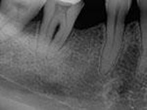
A dental image examination provides an overview of the amount of bone present, indicating the pattern, distribution, and severity of bone loss.
The periapical image is the image of choice for the evaluation of periodontal disease. The paralleling technique is the preferred periapical exposure method for demonstrating anatomic features of periodontal disease as it accurately records the relationship of the height of the crestal bone to the tooth root. Bisected periapical images may appear to show less bone loss than is accurately present.
Images alone cannot be used to diagnose periodontal disease as they do not provide information about the condition of soft tissue or early bone changes.
They are two-dimensional representations of three-dimensional objects:
-
- Buccal and lingual areas may be difficult to evaluate
- Bone loss may be difficult to detect in furcation areas
 |
| Furcation area: The area between the roots of multirooted teeth. |
