34.1
The periodontium refers to the tissues that invest and support the gingiva and alveolar bone.
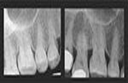
Lamina dura appears as a dense radiopaque line in healthy teeth
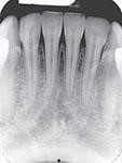
The alveolar crest is about 1.5 to 2 mm apical to the CEJ of adjacent healthy teeth. In anterior teeth, the alveolar crest is pointed and sharp and appears to be very radiopaque. In posterior teeth, the alveolar crest appears flat and smooth, and parallel to a line between adjacent cementoenamel junctions. It appears a little less radiopaque than in anterior teeth.
 |
 |
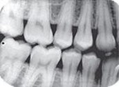 |
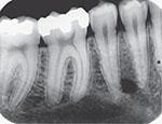
| Healthy alveolar bone on a periapical image | Healthy alveolar crest in anterior region that appears pointed | Healthy alveolar crest in posterior region appears flat, smooth and radiopaque in horizontal BW image |
Periodontal ligament space appears as a thin radiolucent line between the root of the teeth and the lamina dura. It is continuous around the root structure and of uniform thickness in healthy teeth.
Periodontal Disease
Periodontal disease is a group of diseases that affect the tissue around teeth. May range from superficial inflammation of gingiva to destruction of supporting bone and periodontal ligament. The gingiva appears swollen, red, and bleeding, with soft tissue pocket formation. Periodontal disease is most prevalent in adults and is totally preventable with good daily oral self care and regular monitoring appointments with the dental hygienist and/or dentist.
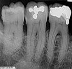
Paralleling technique most accurately will represent level of bone. Bisecting technique can distort the level of bone present due to the vertical angulations.
The image appearance is different: the alveolar crest appears indistinct and bone loss is seen.
 |
 |
| Height of crestal bone is accurately represented by the PA image LCP technique exposed | Bisecting technique distorts the level of bone due to vertical angulations left image and LCP right image. Note the difference in bone level. With paralleling the height of the crestal bone and bone loss is clearly seen. |
Clinical examination including periodontal probing and gingival examination. Whenever clinical evidence is present, a dental image examination must be used in conjunction with a clinical examination to get maximum diagnostic information.
Therefore, detection of periodontal disease generally requires both a clinical and dental image examination. The clinical examination provides information about soft tissue. Clinical examination provides information about soft tissues, whereas dental images permit evaluation of hard tissues, such as bone. The image examination provides information about bone.
